
Page 58 - IJB-10-2
P. 58
International Journal of Bioprinting Advancements in 3D printing
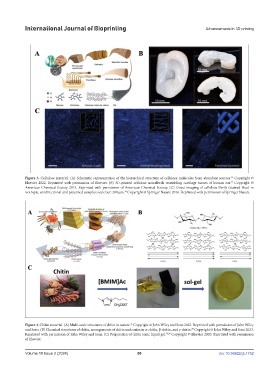
Figure 3. Cellulose material. (A) Schematic representation of the hierarchical structure of cellulose molecules from abundant sources. Copyright ©
32
Elsevier 2022. Reprinted with permission of Elsevier. (B) 3D-printed cellulose nanofibrils resembling cartilage tissues of human ear. Copyright ©
33
American Chemical Society 2015. Reprinted with permission of American Chemical Society. (C) Direct imaging of cellulose fibrils (stained blue) in
isotropic, unidirectional and patterned samples; scale bar: 200 μm. Copyright © Springer Nature 2016. Reprinted with permission of Springer Nature.
34
Figure 4. Chitin material. (A) Multi-scale structures of chitin in nature. Copyright © John Wiley and Sons 2023. Reprinted with permission of John Wiley
35
and Sons. (B) Chemical structures of chitin; arrangements of chitin molecules in α-chitin, β-chitin, and γ-chitin. Copyright © John Wiley and Sons 2023.
35
Reprinted with permission of John Wiley and Sons. (C) Preparation of chitin ionic liquid gel. 35,36 Copyright © Elsevier 2020. Reprinted with permission
of Elsevier.
Volume 10 Issue 2 (2024) 50 doi: 10.36922/ijb.1752