
Page 65 - IJB-3-1
P. 65
3D bioprinting of stem cells and polymer/bioactive glass composite scaffolds for bone tissue engineering
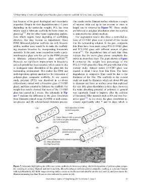
tion because of its good rheological and viscoelastic Fine cracks on the filament surface which are a couple
properties. Despite its slow degradation rate (~2 years of microns wide and up to ten microns or more in
depending on the molecular weight), PCL has been length can be observed in Figure 7C. Those cracks
widely used to fabricate scaffolds for bone tissue en- are believed to aid glass dissolution when the scaffold
gineering [33] . But for other tissue engineering applica- is immersed in the culture medium.
tions which require faster degrading of scaffolding Our degradation results also show a controlled re-
structure, this may become an impediment. Since lease of 13-93B3 glass over a period of two weeks
FDM fabricated polymer scaffolds are only biocom- into the surrounding solution. In the past, composite
patible, another issue would be to make the scaffold- thin films have been made using PCL/13-93B3 glass
ing structure bioactive by incorporating bioceramic and PCL/45S5 glass with different amount of glass
materials. In the past, some researchers made a poly- content [35] . The degradation data of such thin films
mer-bioactive glass wire for use by the FDM process indicate that the entire glass almost completely dis-
to fabricate polymer-bioactive glass scaffolds [34] . solves in about three days. The graph shown in Figure
However, no significant improvement in bioactivity 8 compares the weight loss percentage of the
and cell growth has been reported, which could be due PCL/13-93B3 glass thin films (80 µm) with that of the
to inadequate ionic dissolution of the glass into the current study. Almost entire 13-93B3 glass was
surrounding environment. This makes the FDM and reacted in about 3 da ys from thin films. The faster
melt-deposition options unattractive for fabrication of degradation in composite films could be due to the
polymer-glass composite scaffolds. In our current thickness of the film. The scaffolds in the current
study, polymer (PCL) was dissolved in a s olvent study are made by filaments which are about 400 µm
(chloroform), mixed with a bioactive glass (13-93B3 in diameter and have no surface pores that explained
glass), and then extruded to fabricate the scaffold. Our the very little glass dissolution in three days. However,
weight loss results showed that most of the 13-93B3 the water absorbing potential of polymers in general
glass has reacted in 2 weeks. The schematic in Fig- was reportedly found to improve after the addition
ure 7 explains the difference in the glass dissolution of bioceramic filler materials such as HA and even bio-
from filaments printed using (A) FDM or melt-extrus- active glass [12] . In our study, the glass dissolution in-
ion process and (B) solvent-based extrusion process. creased significantly after 7 and 14 days, which is
Figure 7. Schematic highlighting the difference in two methods of extrusion printing. (A) Melt-deposition of polymer-glass compo-
site resulting in a dense filament and low bioactivity, (B) solvent-based extrusion printed composite resulting in a porous filament
with high bioactivity, (C) SEM images showing surface cracks on the filament indicated by arrows in (i) and (ii), and pores inside the
filament measuring less than 10 µm are also indicated by arrows in (iii).
International Journal of Bioprinting (2017)–Volume 3, Issue 1 61