
Page 101 - IJB-10-3
P. 101
International Journal of Bioprinting 3D bioprinting for vascularized skin tissue engineering
peroxide into hydrogels that were both hypoxic and non- favorable microenvironment for rapid incorporation and
hypoxic. Hydrogels that were hypoxic showed a higher further vascularization. 58-62 Miyazaki et al. developed
63
level of cell independence when compared with those that 3D skin substitutes with vascular networks that did not
were non-hypoxic. Larger cell clusters in both hydrogels require scaffolding. Dermal fibroblasts, vascular ECs, and
showed the recruitment of host cells. In comparison to epidermal keratinocytes were among these cells utilized to
the diphenyleneiodonium chloride (DPI)-treated groups, develop vascularized skin substitutes. In approximately 15
there was a decrease in GFP cell recruitment and cluster days, pre-vascularized 3D skin substitutes developed the
+
area, but there was no significant difference in cluster size desired morphology, and GFP-expressing human umbilical
as shown in Figure 3B–R. vein endothelial cells (HUVECs) showed a significantly
54
Common in all cases, a hypoxic microenvironment high level of viability as well as an effective vasculature with
is an essential component wherein ECs interact to a minimal amount of non-viable cells. Vascular network
form networks of neovessels. By creating a regulated density and branching, vessel formation, and vascular
microenvironment to investigate clustered vasculogenesis, area were all influenced by the optimum cell density
we observed the formation of clusters both in vitro and ratio of HUVECs to FN-G-coated normal human dermal
in vivo within an equivalent timeframe, which allowed fibroblasts (NHDFs). The incorporation of keratinocytes
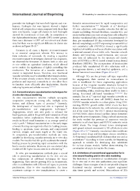
us to confirm the significance of tightly controlling this produced fully vascularized 3D skin substitutes with a
mechanism. The formation of a vascular network is homogeneous dermis, a well-stratified epidermis, and a
crucial in implanted tissues. Therefore, new functional dermal lumenized vasculature (Figure 4A-a–d). 63
vascular networks must be identified after transplantation. Although ECs are the primary cell type responsible
If the structure already contains blood vessels, increased for angiogenesis, their survival in monocultures in
perfusion and improved connectivity in vivo allow them vitro is insufficient for tissue engineering applications
to function more efficiently after implantation, thereby because of the irregular vascular structures formed in
reducing hypoxia and cellular necrosis. 42,47-51,55 monocultures. 49,59,64 Monocultures cause ECs to lose their
3.3. Conventional pre-vascularization techniques for self-assembling ability, rendering them unable to form a
39,49,61,65,66
in vitro skin tissue modeling lasting, functional cell-based vasculature. For
67
In vitro vascularization involves multiple synergistic example, Ren et al. reported high vascular networks on
cooperative components among cells, multiple active human mesenchymal stem cell (hMSC) sheets but no
56
factors, and different types of proteins. Currently, HUVEC vascular networks on culture plates. Using CD31
the development of vascularized skin is improved by staining, HUVEC growth within hMSC sheets has been
pre-vascularization and angiogenesis techniques. demonstrated to be network-like, with networks growing
29
Angiogenesis, which uses skin grafts to enhance the out of the inner layer. The O.C.T. mixture incorporating
healing process, aids in the growth and maturation of host HUVEC/hMSC sheets showed capillary lumen formation
vasculature before implantation. However, this method along with network expansion. Using confocal microscopy,
results in blood vessel formation at an average speed of the study verified the presence of numerous vascular
only 5 µm/h. Delayed vascularization in the early stages networks in cell sheet layers and HUVEC networks inside
13
of wound healing can negatively affect the healing process, hMSC sheets. The image showed lumen development
leading to apoptosis, tissue necrosis, and compromised and upward network movement in 3D cell sheets in vitro
67
nutrient, oxygen, and waste supply at the injury site. (Figure 4B-a–f). Currently, vascularized skin models are
In comparison, pre-vascularization methods are more used to screen drugs and investigate disease conditions.
suitable for developing vascular networks in in vitro skin 3D-bioprinted vascularized full-thickness skin was used as
68
models. Pre-vascularized skin grafts accelerate the wound- a substitute. According to these models, the bioprinting
healing process by providing rapid connections with the approach is suitable for developing skin substitutes
host’s vascular networks. Moreover, pre-vascularized skin for various disease models, further demonstrating the
models exhibit higher success rates in in vitro testing. 57 construction of atopic dermatitis-like tissues.
In vitro, pre-vascularization methods play a crucial 3.4. Limitations of conventional approaches for in
role in promoting vascularization in the skin and are vivo wound healing and in vitro skin modeling
key components of cell-based techniques for tissue The limitations of conventional vascularization strategies
engineering. They involve growing ECs on biomaterials, for in vitro and in vivo skin applications pose significant
often in combination with different cell types, to promote challenges. These strategies often do not provide adequate
proliferation. After in vitro vascularization, the tissue functional vascular networks to support engineered
constructs undergo in vivo transplantation to create a skin tissue growth and survival. Monocultures of ECs
Volume 10 Issue 3 (2024) 93 doi: 10.36922/ijb.1727