
Page 103 - IJB-10-3
P. 103
International Journal of Bioprinting 3D bioprinting for vascularized skin tissue engineering
have exhibited a limited ability to self-assemble into Thus, conventional vascularization strategies for both
proper vascular structures in vitro. The irregularity of in vitro and in vivo skin applications are limited by
these structures hinders the function of the vasculature, the inadequate self-assembly of ECs in monocultures,
compromising the supply of nutrients and oxygen to the slow angiogenesis, and insufficient nutrient delivery to
tissue. The lack of sustained and stable vascularization thicker or avascular tissue implants. Overcoming these
57
in EC monocultures poses a barrier to successful tissue limitations requires innovative approaches such as in vitro
engineering outcomes. Conventional in vivo angiogenesis pre-vascularization techniques to achieve functional and
57
techniques are limited in speed and efficiency. Blood vessels sustained vascularization in engineered skin tissues.
57
are formed slowly, at a rate of approximately 5 μm/h.
Delayed vascularization can slow wound healing, leading 4. 3D bioprinting techniques and bioinks
to apoptosis, tissue necrosis, and insufficient absorption for engineering skin tissue
of nutrients in the wound. Besides, sluggish angiogenesis
makes it challenging to vascularize large tissue constructs 4.1. Potential of 3D skin bioprinting
effectively. Furthermore, the pre-existing vasculature has a The precise deposition of living cells, biomaterials, and
57
theoretical diffusion capacity of approximately 100–200 μm, growth factors in a predefined manner is made possible
which may result in inadequate nutrient and oxygen supply by an advanced additive manufacturing technique known
57
to the central regions of thick or avascular tissue implants. as bioprinting, which uses computer-aided design and
Lack of vascularization can lead to tissue necrosis, employs an layer-by-layer printing process for high
Creating
adaptability and reproducibility (Figure 5).
69,70
compromised functionality, and failure of the implant.
intricate structures that closely resemble the extracellular
To overcome these limitations, researchers have matrix (ECM) using this technique has considerable
explored alternative approaches such as in vitro pre- potential for enhancing cell adhesion and proliferation
vascularization techniques. These methods involve the simultaneously. The benefits of bioprinting include the
cultivation of ECs on biomaterials—often in combination ability to design graded macroscale structures that closely
with other cell types—to promote vascular network resemble the environment in real tissues, encouraging the
formation. Pre-vascularized skin constructs accelerate attachment and growth of various cells. Additionally, the
57
wound healing and improve in vitro testing outcomes. inclusion of microfeatures, such as ridges and modified
57
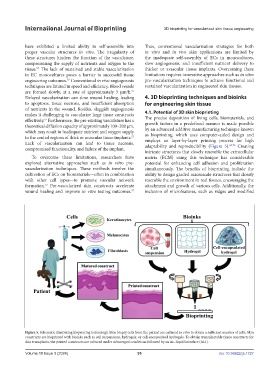
Figure 5. Schematic illustrating bioprinting technology. Skin biopsy cells from the patient are cultured in vitro to obtain a sufficient number of cells. Skin
constructs are bioprinted with bioinks such as cell suspensions, hydrogels, or cell-encapsulated hydrogels. To obtain transplantable tissue constructs for
skin transplants, the printed constructs are cultured under submerged conditions followed by an air–liquid interface (ALI).
Volume 10 Issue 3 (2024) 95 doi: 10.36922/ijb.1727