
Page 98 - IJB-10-3
P. 98
International Journal of Bioprinting 3D bioprinting for vascularized skin tissue engineering
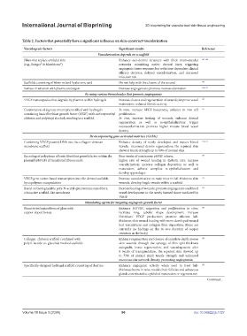
Table 2. Factors that potentially have a significant influence on skin-construct vascularization
Vasculogenic factors Significant results Reference
Vascularization depends on a scaffold
Films that replace artificial skin Enhance neo-dermis structure with thick microvascular 143-148
(e.g., Integra® & Matriderm®) networks mimicking native dermal layer, triggering
angiogenic tissue response, but with time-dependent clinical
efficacy decrease, delayed vascularization, and increased
infection risk
Scaffolds consisting of fibrin or/and hyaluronic acid Do not help with the closure of the wound 149
Surface irradiation with plasma and argon Increase angiogenesis; promote neovascularization 146,150
By using various biomolecules that promote angiogenesis
VEGF nanocapsules that degrade by plasmin within hydrogels Increase closure and regeneration of wounds; improve vessel 149
maturation; reduced fibrotic activity
Combination of alginate microspheres filled with hydrogels In vitro, increase bFGF bioactivity; enhance in vivo cell 151
containing basic fibroblast growth factor (bFGF) with carboxymethyl proliferation
chitosan and polyvinyl alcohol, resulting in a scaffold In vivo, increase healing of wounds; enhance dermal
regeneration as well as re-epithelialization; trigger
neovascularization; promote higher mature blood vessel
density
By incorporating gene-activated matrices (GAMs)
Combining VEGF plasmid DNA into the collagen–chitosan Enhance density of newly developed and mature blood 152,153
membrane scaffold vessels; increased dermis regeneration; the repaired skin
showed tensile strength up to 80% of normal skin
Encoding of polyplexes of basic fibroblast growth factor within the Four weeks of continuous pbFGF release; 154
plasmid (pbFGF) of transfected fibrous mats higher rate of wound healing in diabetic rats; increase
vascularization; increase collagen deposition as well as
maturation; achieve complete re-epithelialization and
develop appendages
VEGF gene vector-based transcription into the dermal scaffolds Increase vascularization in nude mice in full-thickness skin 155
by copolymer encapsulation wounds; develop fragile vessels within a scaffold
Based on biodegradable poly-N-acetyl-glucosamine nanofibers, Increase healing of wounds; promote angiogenesis and blood 156
a bioactive scaffold-like membrane vessel development in the newly formed tissue mediated by
VEGF.
Stimulating agents for targeting angiogenic growth factor
Bioactivated microfibers of glass with Enhance HUVEC migration and proliferation in vitro; 157
copper-doped borate facilitate long, tubular shape development; increase
fibroblasts’ VEGF production; promote efficient full-
thickness skin wound healing with more developed wound
bed vasculatures and collagen fiber deposition; (there are
currently no findings on the in vivo duration of copper
retention in the body)
Collagen–chitosan scaffold combined with Enhance regeneration and closure of complete-depth mouse 158
poly L-lactide-co-glycolide meshed scaffolds skin wounds through the synergy of thin split-thickness
autografts, tissue regeneration, and vasculogenesis; after
8 weeks of transplantation, the repaired skin showed up
to 73% of normal skin’s tensile strength and enhanced
microvascular network density, promoting angiogenesis
Specifically designed hydrogel scaffold consisting of dextran Enhance angiogenic activity when used to treat full- 159
thickness burns in mice models; hair follicles and sebaceous
glands contributed to epithelial maturation or regeneration
Continued...
Volume 10 Issue 3 (2024) 90 doi: 10.36922/ijb.1727