
Page 102 - IJB-10-3
P. 102
International Journal of Bioprinting 3D bioprinting for vascularized skin tissue engineering
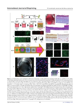
Figure 4. (A) Biofabrication of pre-vascularized scaffold-free 3D skin tissue. (a) Main method for producing vascular skin as a substitute in vitro; HEKn:
neonatal human epidermal keratinocytes. (b) In vitro vascular network development in dermo-epidermal 3D skin replacements. HUVEC-expressing cells were
co-cultured with normal human dermal fibroblasts (NHDF) and grown for up to 7 days before being covered with HEKn cells. (c) The 3D skin replacements
developed an interconnected vascular network in 7 days because of epidermal differentiation. FN-G-coated NHDF was supported, while HUVECs were
5
seeded at 0.1–1 × 10 cells per culture. Co-culturing of 1000:1, 500:1, or 100:1 FN-G-coated NHDFs with HUVECs was performed, followed by the addition of
HEKn. (d) In vitro evaluation of 3D skin replacements with pre-vascularization was performed using HUVECs mixed with FN-G-coated NHDF and HEKn,
63
which were maintained for up to 7 days. Figure 4A was reprinted from (Copyright © 2019, with permission from Springer Nature). (B) (a) Schematic of 3D
pre-vascularized skin replacement construction in the in vitro condition, showing the process and the morphology of human mesenchymal stem cells (hMSCs)
after 14 days. (b) (i) Confluence was achieved, resulting in a thick cell layer. Fluorescent images ((ii)–(vi)) indicated the development of HUVEC networks on
an hMSCs sheet after HUVECs were sown there for 2 h (ii), 1 day (iii), and 7 days (iv). The shape and size of HUVECs grown on tissue culture plates after 2
h (v) and 7 days (vi) are shown in fluorescent images. On the tissue culture plates, HUVECs proliferated but were not arranged into networks (scale bar: 100
μm). (c) A cell sheet in its entirety (scale bar: 3.4 mm). Following 7 days of growth, confocal microscopy images showed the development of HUVEC lumens
on human mesenchymal stem cell (hMSC) sheets. Lumens developed on the hMSCs sheet (d) (scale bar: 100 µm) and moved within the surface of the sheet
67
(e) (cross-sectional view; scale bar: 50 µm), as revealed by CD31 immunofluorescent staining images. When a single cell sheet was folded into a 3D pre-
vascularized construct, it was cryosectioned (f), and CD31 staining revealed that the structure had developed networks and lumens (g) (white arrows; scale
67
bar: 100 μm). Figure 4B-b–g were reprinted from (Copyright © 2014, with permission from Hindawi).
Volume 10 Issue 3 (2024) 94 doi: 10.36922/ijb.1727