
Page 99 - IJB-10-3
P. 99
International Journal of Bioprinting 3D bioprinting for vascularized skin tissue engineering
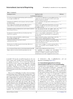
Table 2. Continued...
Vasculogenic factors Significant results Reference
By cell-mediated angiogenesis
3D constructions integrating fetal fibroblasts with human dermal Third- and second-degree burns in young children heal more 160
microvascular endothelial cells rapidly without the use of autografts; excellent aesthetic and
functional results were achieved
Endothelial cells (HUVECs) within hyaluronic acid-based scaffolds Increase endothelial cell organization and development of 161
for artificial dermis micro-capillaries
Use of collagen with and without glycosaminoglycan in the co- Enhance in vitro capillary-like tube generation and increase 162-164
culture of HUVECs with fibroblasts and/or keratinocytes formation of tubular structures interconnected with mesh-
like capillary networks
Endothelial progenitor cell administration can be achieved with both Cells were incorporated into the capillary wall at the 165-168
local and systemic delivery location of the injury as well as by ischemia; contractile and
vasomotor activity could be seen in the developing vessels.
Incorporation of endothelial cells into a dermal construct from Achieve a more effective epithelialization of the matrix 169
human blood outgrowth structure and effective revascularization and oxygenation of
the wound bed
Micro-tissues constructed with fibrin, seeded by human Higher endothelial sprouting occurs in larger vessels that 170
microvascular endothelial cells spread out across a substantial distance (1–2 mm)
Conditioned medium for bone marrow-derived Enhance migration and proliferation of endothelial cells in 171
mesenchymal stem cells culture; local injections increased collateral perfusion and
improved limb function in vivo
Biofabrication of scaffolds using mesenchymal stem cells derived Improve repair or healing of wounds by cutaneous, fibrous, 172-174
from within the bone marrow adipose, and especially vascular-endothelium tissues in
mouse models
In vivo treatment of human adipose-derived mesenchymal stem cells A higher density of microvascular tissue was seen 175
Human umbilical vein blood-derived mesenchymal stem cells The engraftment of umbilical cord blood-derived 176-178
mesenchymal stem cells (UCB-MSC) in a mouse model with
an ischemic hind limb increased the regenerative ability of
skeletal muscles, but its role in angiogenesis remains unclear
is needed. During the wound-healing process, the new of inflammatory cells), re-epithelialization, and scar
34
blood vessels must develop from pre-existing ones by deposition/remodeling (Figure 2B). 40
an angiogenesis process. ECs at vessel tips initiate the
35
development of new vessels during angiogenic sprouting 3.2. Conventional approaches to enhance
by degrading the interstitial proteins and basement vascularization of skin for in vivo wound healing
membrane, resulting in branching. While the role of Increased levels of proangiogenic factors in transplanted
34
angiogenesis in the healing of wounds in mature skin has cells and physiological inflammation—particularly in the
been extensively studied, that of vasculogenesis remains up hypoxic environment of wounds—induce in vivo tissue
for debate. 33 engineering transplantation, promoting vascularization
However, it is challenging to develop a microvasculature during wound healing. In the absence of a fully functional
network in large tissue substitutes after transplantation. For vasculature, cells seeded or encapsulated in bioink in
36
successful vascularization, the physiology of cells and viability the 3D construct develop hypoxic environments owing
after transplantation are essential for the formation of new to insufficient nutrient supply. Blood vessel formation
blood vessels. Tissue substitutes with a general size of 0.1 to was observed in the surrounding host tissue growing
10 cm experience oxygen and nutrition deficits compared in a 3D-bioprinted construct. 38,39 The biological process
to other existing vessels with a size of 100 to 200 μm. This of angiogenesis occurs slowly, incorporating tens of
sizable difference causes cell death along with the failure of micrometers of tissue each day. Large implants require an
the implant. 37-39 Considering effective vascularization both extended period for vascularization because of this delayed
in vitro and in vivo, it is essential to comprehend three stages process, which can lead to the functional and structural loss
of human skin wound healing including immediate injury of nutrient-starved cells and eventual cell death. However,
responses (clot formation), inflammation (recruitment in thin or avascular tissue implants, this timeframe may
Volume 10 Issue 3 (2024) 91 doi: 10.36922/ijb.1727