
Page 96 - IJB-10-3
P. 96
International Journal of Bioprinting 3D bioprinting for vascularized skin tissue engineering
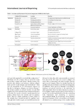
Table 1. Locations and functions of structural components of different skin layers
Cellular/ECM components Location Function
Epidermis Keratinocyte Entire layers As component of skin boundary, synthetic keratin
Melanocyte Stratum germinativum For synthesis of melanosomes
Merkel cell Stratum germinativum As sensor
Langerhans cell Spinous layer/prickle cell layer/ Present antigens to the immune system
distinctive keratinocytes
Laminin Basement membrane Connect epidermis and dermis
Dermis Fibroblast Entire dermis layer Synthesize ECM, secrete cytokines, and
promote wound healing
Collagen fiber Entire dermis layer For support and protection
Blood vessel Dermis of papillaries Maintain the skin’s oxygen and nutrient levels
Elastic fiber Reticular dermis For the elasticity of skin
Nerve Venules and arterioles Enable various sensations associated with touch, pressure,
temperature, and itching
Hair follicle Dermis reticular Form physical barrier, inhibit scar formation, and for
antibacterial effect
Sweat gland Dermis reticular Maintain body temperature
Figure 1. Schematic of the structure and function of human skin.
and mast cells organized in a network-like configuration. delivered to skin cells, while waste materials are removed
23
Fibroblasts are the primary cells in connective tissue from the dermis. This layer also contains collagen and
that produce collagen and elastin. Elastin proteins help elastin fibers, contributing to the skin’s strength, elasticity,
24
maintain skin elasticity and resilience, whereas collagen and structural integrity. Above the reticular layer lies the
fibers provide tensile strength and stretchability. Mast papillary layer, which features a more delicate arrangement
25
cells are responsible for the inflammatory response of of capillaries. These capillaries stimulate immunological
the skin to pathogens, allergens, and physical damage. responses and provide nutrition to the epidermis, the
26
The dermis includes an intricate vascular structure that skin’s upper layer. These two layers function in concert to
supplies nutrients to maintain its homeostasis and function form a highly complex vascular network whose function is
of the skin. The vascular network is mainly located at two essential for maintaining the health of the skin, controlling
primary dermal layers: the reticular and the papillary body temperature, and responding to numerous
layers. There is a dense meshwork of blood vessels in environmental stimuli. In dermatology, wound healing,
the reticular layer, located beneath the papillary layer. and the development of skin care products and therapies
Through these vessels, oxygen and essential nutrients are all depend on an understanding of the complex dermal
Volume 10 Issue 3 (2024) 88 doi: 10.36922/ijb.1727