
Page 554 - IJB-10-3
P. 554
International Journal of Bioprinting Engineered 3D-printed PVA vascular grafts
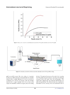
Figure 9. Stress–strain curves for unmodified poly(vinyl alcohol) (PVA) grafts and biofunctionalized PVA grafts.
Figure 10. Schematic representation of biofunctionalized poly(vinyl alcohol) (PVA) graft flow testing.
graft successfully passed the test, owing to its greater 60 min. Throughout the process, the water level covering
mechanical stability. The input was facilitated by a metallic the PVA graft remained constant, indicating that drainage
channel with a smaller diameter (0.3 mm) to maintain a occurred solely with water from the feeding glass. This
continuous flow without fluctuations, while the metallic observation suggests that any potential fluid leakage was
channel with a larger diameter (0.8 mm) served as the effectively prevented. The collected water was measured to
output. The fluid flow consisted of eight cycles, each lasting determine the flow velocity, which was calculated to be 11
Volume 10 Issue 3 (2024) 546 doi: 10.36922/ijb.2193