
Page 555 - IJB-10-3
P. 555
International Journal of Bioprinting Engineered 3D-printed PVA vascular grafts
cm/s. These results align well with the reported conditions cells normally present in the outer layer of the vascular
of normal vasculature documented in the literature. 64 wall; and (iii) U937 monocyte-like cells to represent
immune cells.
3.9. Biological assessment
Vascular graft biocompatibility in the vascular tissue 3.9.1. Biocompatibility assessment of initial vascular
environment is an essential requirement for newly grafts using the extract method
developed materials to be used for such applications. The The biocompatibility of the initial vascular graft (unmodified
65
biological behavior of the vascular graft was investigated and 3D-3H) for endothelial cells was tested using the
in vitro for cells present in the natural vascular tissue. For extract method, according to ISO 10993-5:2009 and ISO
this, we employed (i) bEnd.3 endothelial cells to represent 10993-12:2012. The extract medium was diluted to 1:1, 1:2,
cells in the lumen; (ii) L929 to represent fibroblast 1:5, and 1:10, respectively. The viability of the endothelial
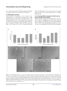
Figure 11. Biocompatibility assessment of initial vascular grafts using the extract method. (A) Viability of bEnd.3 endothelial cells following a 24-h
incubation with vascular graft extract and dilutions of the extract in cell culture medium (via MTT assay). Data presented as mean ± standard deviation;
*p < 0.05; ***p < 0.001. (B) Levels of lactate dehydrogenase (LDH) released from bEnd.3 endothelial cells following a 24-h incubation with vascular graft
extract and dilutions of the extract in cell culture medium (via LDH assay). Data presented as mean ± standard deviation; *p < 0.05; **p < 0.01. (C–G)
Morphology of bEend.3 endothelial cells following a 24-h incubation with vascular graft extract and dilutions of the extract in cell culture medium: (C)
negative control (NC), (D) 1:1 graft extract, (E) 1:2 graft extract in cell culture medium, (F) 1:5 graft extract in cell culture medium, and (G) 1:10 graft
extract in cell culture medium. Magnification: ×10.
Volume 10 Issue 3 (2024) 547 doi: 10.36922/ijb.2193