
Page 558 - IJB-10-3
P. 558
International Journal of Bioprinting Engineered 3D-printed PVA vascular grafts
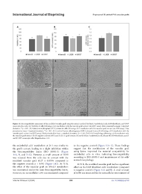
Figure 13. Biocompatibility assessment of the modified vascular graft using the extract method for bEnd.3 endothelial cells, L929 fibroblasts, and U937
monocyte-like cells. (A) Viability of cells following a 24-h incubation with the vascular graft extract (via MTT assay). Data presented as mean ± standard
deviation; **p < 0.01. (B) Adenosine triphosphate (ATP) content in cells following a 24-h incubation with the vascular graft extract (via ATP assay). Data
presented as mean ± standard deviation; **p < 0.01. (C) Levels of lactate dehydrogenase (LDH) released from cells following a 24-h incubation with the
vascular graft extract (via LDH assay). Data presented as mean ± standard deviation; *p < 0.05. (D–I) Cell morphology following a 24-h incubation with
the vascular graft extract: (D–F) negative controls (NC) and (G–I) 1:1 graft extract; (D and G) bEnd.3 endothelial cells, (E and H) L929 fibroblasts, and (F
and I) U937 monocyte cells. Magnification: ×10.
the endothelial cells’ metabolism at 24 h was similar to to the negative control (Figure 15A–C). These findings
the graft’s extract, leading to a slight inhibition within suggest that the modification of the vascular graft
the biocompatibility limits (ISO 10993-5) (Figure using lysine improved the material compatibility for
14A, B, and D–G). However, a small amount of LDH endothelial cells in vitro, indicating biocompatibility
was released from the cells due to contact with the according to ISO-10993-5 and maintenance of the cells’
modified vascular graft (0.27 ± 0.033% compared to normal morphology.
the negative control; p < 0.05) (Figure 14C). At 72 h, At 24 h, the modified vascular graft had no significant
the effect of the vascular graft on bEnd.3 metabolism effect on the L929 fibroblast cells’ metabolism (compared
was maintained under the same biocompatibility limits. to negative control) (Figure 14A and B). A small amount
However, no extracellular LDH was measured compared of LDH was measured in the extracellular environment of
Volume 10 Issue 3 (2024) 550 doi: 10.36922/ijb.2193