
Page 65 - IJB-4-1
P. 65
An nMgO containing scaffold: Antibacterial activity, degradation properties and cell responses
differentiation by SEM observation, CCK-8 assay and Co., Ltd., China), followed by observing with light
ALP staining, respectively. microscope.
For evaluating cellular adhesion, the cell/scaffold
specimens were gently washed with PBS, followed 2.7 Statistical Analysis
by fixing with 2.5% glutaraldehyde. Subsequently, a Quantitative data were expressed as the mean ± standard
graded ethanol series was used to dehydrate the cells. deviation. Levene’s test was applied to examine equality
Afterwards, the specimens were dried in vacuum drying of variances. Unpaired two-tailed Student’s t-test was
oven, followed by sputtering with platinum. Finally, the performed to determine statistical significance. Labels *,
cellular morphologies were characterized by Phenom ** and *** represent p < 0.05, p < 0.01 and p < 0.001,
ProX SEM using backscattering mode under 15 kV respectively.
acceleration voltage. For CCK-8 assay, the MG63 cells
were harvested from the scaffold specimens by Trypsin- 3. Results and Discussion
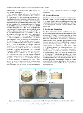
ethylene diamine tetraacetic acid solution, followed The three-dimensional porous scaffold model and a
by centrifugation treatment. Afterwards, 0.2 mL of representative SLS-fabricated PHBV/nMgO scaffold
the obtained supernatant was added into 1 mL of fresh
culture medium, followed by adding CCK-8 (CK04- were shown in Figure 1. It could be seen that the shape
13, Dojindo Molecular Technologies, Kimamoto, and size of the scaffold were consistent with those of
Japan) solution into it according to the instructions of the model. The scaffold showed a well ordered and
the manufacturer, which generated an orange formazan interconnected porous structure. It was considered that
product by cellular dehydrogenases. After culture for 4 h the pore size of scaffolds should be large enough to
at 37 °C, 100 μL of the sample solution was transferred ensure nutrient delivery and tissue ingrowth but not too
[30]
[31]
into plate to measure the absorbance with a microplate large to prevent cell migration . Roosa et al. found
reader (Beckman, USA) at 450 nm. For ALP staining, all of the polycaprolactone scaffolds with pore size
the MG63 cells were washed with PBS, fixed with from 350 to 800 μm could promote bone regeneration
4% paraformaldehyde for 30 min and then permeated and there were no significant differences in new bone
with 0.5% Triton X-100 for 30 min. Subsequently, formation between them. Similar results were reported
ALP staining was carried out with 5-bromo-4-chloro- by Schek et al. [32] who found significant new bone
3-indolyl-phosphate/NBT (BCIP/NBT) alkaline formation for both poly(propylenefumarate)/β-tricalcium
phosphatase color development kit (P0321, Shanghai phosphate scaffolds with 300 and 800 μm pores, with no
Beyotime Biological Technology Co., Ltd, China) statistical differences between them. Hence, the PHBV/
according to the instructions of the manufacturer. nMgO scaffold with pore size of about 400 μm may be
Finally, the stained cells were mounted with water- beneficial for substance metabolism, cell responses and
based mounting medium (Boster Biological Technology bone regeneration.
(A) (B) (C)
(D) (E) (F)
Figure 1. (A–C) The three-dimensional porous scaffold model and (D–F) a representative SLS-fabricated PHBV/nMgO scaffold.
4 International Journal of Bioprinting (2018)–Volume 4, Issue 1