
Page 69 - IJB-4-1
P. 69
An nMgO containing scaffold: Antibacterial activity, degradation properties and cell responses
they would compel them generate oxidative stress, that for the PHBV scaffolds. This was mainly ascribed
which subsequently led to the damage of the structure to the alkaline degradation products of MgO, which
and functions of bacteria [51,52] . Besides, the contact exerted neutralization effect against the acid degradation
action of the MgO nanoparticles on bacteria would products of PHBV. These results indicated nMgO could
make them generate mechanical stress, resulting in the promote the degradation of the PHBV scaffolds and
deformation and damage of the bacterial structure [53,54] . neutralize their acid degradation products.
In addition, there were large amounts of active sites The surface microtopography of PHBV/5%nMgO and
on MgO nanoparticles [55] , enabling them easily absorb PHBV scaffolds after immersion were characterized by
to the bacteria; the enrichment of nanoparticles on the SEM (Figure 9) to explain the results of mass loss and
[56]
bacteria would increase their membrane permeability . pH. It was clear that the surface morphologies of the
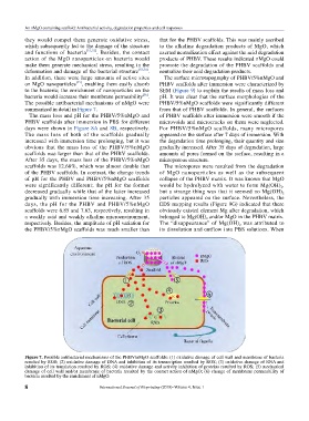
The possible antibacterial mechanisms of nMgO were PHBV/5%nMgO scaffolds were significantly different
summarized in detail in Figure 7. from that of PHBV scaffolds. In general, the surfaces
The mass loss and pH for the PHBV/5%nMgO and of PHBV scaffolds after immersion were smooth if the
PHBV scaffolds after immersion in PBS for different microvoids and microcracks on them were neglected.
days were shown in Figure 8A and 8B, respectively. For PHBV/5%nMgO scaffolds, many micropores
The mass loss of both of the scaffolds gradually appeared on the surface after 7 days of immersion. With
increased with immersion time prolonging, but it was the degradation time prolonging, their quantity and size
obvious that the mass loss of the PHBV/5%nMgO gradually increased. After 35 days of degradation, large
scaffolds was larger than that of the PHBV scaffolds. amounts of pores formed on the surface, resulting in a
After 35 days, the mass loss of the PHBV/5%nMgO microporous structure.
scaffolds was 12.68%, which was almost double that The micropores were resulted from the degradation
of the PHBV scaffolds. In contrast, the change trends of MgO nanoparticles as well as the subsequent
of pH for the PHBV and PHBV/5%nMgO scaffolds collapse of the PHBV matrix. It was known that MgO
were significantly different; the pH for the former would be hydrolyzed with water to form Mg(OH) 2 ,
decreased gradually while that of the latter increased but a strange thing was that it seemed no Mg(OH)
2
gradually with immersion time increasing. After 35 particles appeared on the surface. Nevertheless, the
days, the pH for the PHBV and PHBV/5%nMgO EDS mapping results (Figure 9G) indicated that there
scaffolds were 6.85 and 7.63, respectively, resulting in obviously existed element Mg after degradation, which
a weakly acid and weakly alkaline microenvironment, belonged to Mg(OH) and/or MgO in the PHBV matrix.
2
respectively. Besides, the amplitude of pH variation for The “disappearance” of Mg(OH) was attributed to
2
the PHBV/5%nMgO scaffolds was much smaller than its dissolution and outflow into PBS solutions. When
Figure 7. Possible antibacterial mechanisms of the PHBV/nMgO scaffolds: (1) oxidative damage of cell wall and membrane of bacteria
resulted by ROS; (2) oxidative damage of DNA and inhibition of its transcription resulted by ROS; (3) oxidative damage of RNA and
inhibition of its translation resulted by ROS; (4) oxidative damage and activity inhibition of proteins resulted by ROS; (5) mechanical
damage of cell wall and/or membrane of bacteria resulted by the contact action of nMgO; (6) change of membrane permeability of
bacteria resulted by the enrichment of nMgO.
8 International Journal of Bioprinting (2018)–Volume 4, Issue 1