
Page 566 - IJB-10-4
P. 566
International Journal of Bioprinting Light-based muscle bioprinting with bioglass
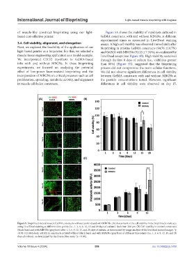
of muscle-like construct bioprinting using our light- Figure 5A shows the viability of myoblasts cultured in
based cost-effective printer. GelMA constructs, with and without MBGNs, at different
experimental times as measured in Live/Dead staining
3.4. Cell viability, alignment, and elongation assays. A high cell viability was observed immediately after
Next, we explored the feasibility of the application of our bioprinting in pristine GelMA constructs (94.76 ±10.7%)
light-based printer as a bioprinter. For this, we selected a and GelMA with MBGNs (91.25 ± 7.34%), as confirmed by
muscle tissue engineering application as a model example. Live/Dead assays (see Figure 5B). High viability, sustained
We incorporated C2C12 myoblasts to GelMA-based through the first 8 days of culture (i.e., viabilities greater
inks with and without MBGNs. In these bioprinting than 80%) (Figure 5B), suggested that the bioprinting
experiments, we focused on analyzing the potential process did not compromise the main cellular functions.
effect of low-power laser-assisted bioprinting and the We did not observe significant differences in cell viability
incorporation of MBGNs on critical processes such as cell between GelMA constructs with and without MBGNs at
proliferation, spreading, metabolic activity, and alignment the particle concentrations tested. However, significant
in muscle cell-laden constructs. differences in cell viability were observed on day 15,
Figure 5. Bioprinted skeletal muscle GelMA constructs without (control) and with MBGNs. (A) Assessment of the cell viability in the bioprinted constructs
using Live/Dead staining at different time points (i.e., 1, 3, 6, 9, 12, 15 and 20 days of culture). Scale bar: 200 μm. (B) Cell viability in control constructs
(black bars) and with MBGNs (gray bars) after 1, 3, 6, 9, 12, 15, and 20 days of culture, as determined by image analysis of the live/dead stained images. *p
<0.05. (C) Metabolic activity in constructs printed without (black bars) and with MBGNs (gray bars) at different time points (i.e., 1, 3, 6, 9, 12, 15, and 20
days of culture), as determined by the Presto Blue assay. *p < 0.001.
Volume 10 Issue 4 (2024) 558 doi: 10.36922/ijb.1830