
Page 568 - IJB-10-4
P. 568
International Journal of Bioprinting Light-based muscle bioprinting with bioglass
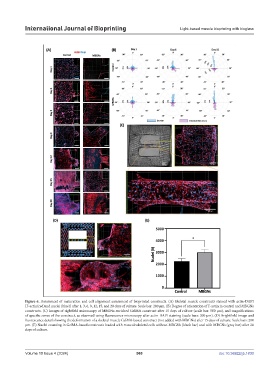
Figure 6. Assessment of maturation and cell alignment assessment of bioprinted constructs. (A) Skeletal muscle constructs stained with actin-DAPI
[F-actin(red)and nuclei (blue)] after 1, 3, 6, 9, 12, 15, and 20 days of culture. Scale bar: 200 μm. (B) Degree of orientation of F-actin in control and MBGNs
constructs. (C) Images of rightfield microscopy of MBGNs-enriched GelMA construct after 15 days of culture (scale bar: 500 μm), and magnifications
of specific zones of the construct, as observed using fluorescence microscopy after actin-DAPI staining (scale bars: 200 μm). (D) Brightfield image and
fluorescence detail showing the deformation of a skeletal muscle GelMA-based construct (not added with MBGNs) after 15 days of culture. Scale bars: 200
μm. (E) Nuclei counting in GelMA-based constructs loaded with musculoskeletal cells without MBGNs (black bar) and with MBGNs (gray bar) after 20
days of culture.
Volume 10 Issue 4 (2024) 560 doi: 10.36922/ijb.1830