
Page 28 - IJB-5-1
P. 28
Shuai C
EBM was first developed and patented by Swedish As a permanent implant, the Ti-6Al-4V scaffolds with
Arcam Company [142] . The EBM equipment is mainly high anti-corrosion ability led to reduced precipitate of
composed of an electron beam gun compartment and a harmful metallic ion, such as Al and V ions, which might
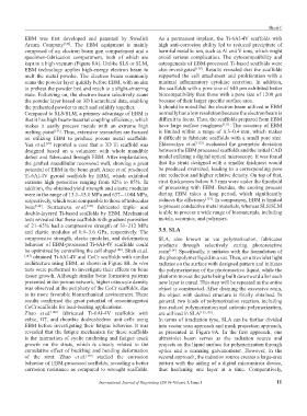
specimen-fabrication compartment, both of which are avoid serious complication. The cytocompatibility and
kept in a high vacuum (Figure 8A). Unlike SLS or SLM, osteogenesis of EBM-processed Ti-based scaffolds were
EBM technology applies high-energy electron beam to also investigated [150] . Results revealed that the scaffolds
melt the metal powder. The electron beam commonly supported the cell attachment and proliferation with a
scans the powder layer quickly before EBM, with an aim minimal inflammatory cytokine secretion. In addition,
to preheat the powder bed and reach to a slight-sintering the scaffolds with a pore size of 640 μm exhibited better
state. Following on, the electron beam selectively scans biocompatibility than those with a pore size of 1200 μm
the powder layer based on 3D hierarchical data, enabling because of their larger specific surface area.
the preheated powder to melt and solidify together. It should be noted that the electron beam utilized in EBM
Compared to SLS/SLM, a primary advantage of EBM is normally has a low resolution because the electron beam is
that it has high beam-material coupling efficiency, which difficult to focus. Thus, the scaffolds prepared from EBM
makes it easily process metals with an extreme high have large surface roughness [151] . The accuracy of EBM
melting point [143] . Thus, extensive researches are focused is limited within a range of 0.3–0.4 mm, which makes
on utilizing EBM to produce porous metal scaffolds. it difficult to fabricate scaffolds with a small pore size.
[29]
Yan et al. reported a case that a 3D Ti scaffold was Eldesoukya et al. [152] evaluated the geometric deviation
designed based on a volunteer with whole mandible between the EBM processed scaffolds and the initial CAD
defect and fabricated through EBM. After implantation, model utilizing a digital optical microscope. It was found
the grafted mandibular recovered well, showing a great that the struts designed with a smaller thickness would
potential of EBM in the bone graft. Ataee et al. produced be produced oversized, leading to a corresponding pore
Ti-6Al-4V gyroid scaffolds by EBM, which exhibited size reduction and higher relative density. On top of that,
extreme high porosities ranging from 82% to 85%. In strut thicknesses below 0.5 mm were under the threshold
addition, the obtained yield strength and elastic modulus of processing with EBM. Besides, the cooling process
were in the range of 13.1–15.0 MPa and 637–-1084 MPa, during EBM takes a long period, which significantly
respectively, which were comparable to those of trabecular reduces the efficiency [153] . In comparison, EBM is limited
bone [144] . Surmeneva et al. [145] fabricated triple- and to process conductive metal materials, whereas SLS/SLM
double-layered Ti-based scaffolds by EBM. Mechanical is able to process a wide range of biomaterials, including
tests revealed that these scaffolds with gradient porosities metals, ceramics, and polymers.
of 21–65% had a compressive strength of 31–212 MPa
and elastic modulus of 0.9–3.6 GPa, respectively. The 3.5. SLA
compressive strength, elastic modulus, and deformation SLA, also known as vat polymerization, fabricates
behavior of EBM-processed Ti-6Al-4V scaffolds could products through selectively curing photoreactive
be optimized by controlling the cell shape [146] . Shah et al. resin [154] . Specifically, it initiates with the formulation of
[147] obtained Ti-6Al-4V and CoCr scaffolds with similar the photopolymer liquid in a vat. Then, an ultraviolet light
architecture using EBM, as shown in Figure 8B. In vivo radiates on the surface with designed pattern and initiates
tests were performed to investigate their effects on bone the polymerization of the photoreactive liquid, while the
tissue growth. Although similar bone formation patterns platform moves the parts being built downward after each
presented in the porous network, higher osteocyte density new layer is cured. This step will be repeated as the entire
was observed at the periphery of the CoCr scaffolds, due object is constructed. After draining the excessive resin,
to its more favorable biomechanical environment. These the object with desired structure is finally obtained. In
results confirmed the great potential of osseointegrated general, two kinds of polymerization reaction, including
CoCr scaffolds for load-bearing applications. free-radical polymerization and cationic polymerization,
Zhao et al. [148] fabricated Ti-6Al-4V scaffolds with are utilized in SLA [155,156] .
cubic, G7, and rhombic dodecahedron unit cells using In terms of irradiation type, SLA can be further divided
EBM before investigating their fatigue behavior. It was into vector scan approach and mask projection approach,
revealed that the fatigue mechanism for these scaffolds as presented in Figure 9A. In the first approach, one
is the interaction of cyclic ratcheting and fatigue crack ultraviolet beam serves as the radiation source and
growth on the struts, which is closely related to the projects on the liquid surface for polymerization through
cumulative effect of buckling and bending deformation optics and a scanning galvanometer. However, in the
of the strut. Zhao et al. [149] studied the corrosion second approach, the radiation source creates a large-area
behavior of EBM-processed scaffolds, revealing a better pattern with the aiding of a digital micromirror device,
corrosion resistance as compared to wrought scaffolds. thus hardening one layer at a time. Comparatively,
International Journal of Bioprinting (2019)–Volume 5, Issue 1 11