
Page 29 - IJB-5-1
P. 29
Additive manufacturing of bone scaffolds
A B
C D
E
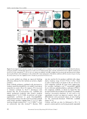
Figure 9. (A) A diagram for two irradiation types for stereolithography (SLA), including vector scan and mask projection. (B) Poly(trimethylene
carbonate) (PTMC) scaffolds fabricated by SLA with various hydroxyapatite (HA) contents, with PTMC20 and PTMC40 containing 20
and 40 wt.% HA, respectively [160] . (C) In vitro cell culture on scaffolds, with (D) scanning electron microscope and fluorescence images
showing the different cell morphologies on the scaffold. (E) Contact radiographs of the defects combined with fluorescence images showing
the newly formed bone after implantation for 2 weeks (in green) and 4 weeks (in red).
the second method can obtain an improved building was also used in SLA to produce scaffolds with a large
efficiency, whereas the first method can achieve a higher elastic modulus range (5.3±0.9–74.6±1.5 kPa). There
accuracy. are also a small number of reports on the use of SLA to
Some synthetic polymers combined with photoreactive build composite scaffolds for bone tissue engineering.
features, good biocompatibility, and suitable mechanical For example, Guillaume et al. [160] successfully applied
properties are used in SLA. For instance, PCL was used SLA to fabricate poly(trimethylene carbonate) (PTMC)/
to fabricate scaffolds by SLA [157] . Mechanical tests HA composite scaffolds, as shown in Figure 9B. The
showed that the SLA-processed PCL scaffolds had incorporated HA was enriched on the surface of scaffolds,
elastic mechanical properties with Young’s modulus forming a microscale structured. In vitro and in vivo
ranging from 6.7 to 15.4 MPa. In addition, cell culture experiments revealed an improved marrow stem cell
experiments confirmed its good biocompatibility. differentiation and accelerated kinetic of bone healing
Poly(tetrahydrofuran) was also used for printed scaffolds for the microscale-structured PTMC/HA scaffolds
with Young’s modulus ranging from 5.7 to 27.5 MPa, (Figure 9C-E).
bending strength ranging from 1.1 to 3.5 MPa [158] , where Ceramic scaffolds can also be fabricated by SLA. In
no cytotoxicity was also showed [159] . Besides, PEG general, ceramic particles are homogeneously suspended
12 International Journal of Bioprinting (2019)–Volume 5, Issue 1