
Page 24 - IJB-5-1
P. 24
Shuai C
[89]
Nevertheless, further investigations using in vitro and photon polymerization , also demonstrate great potential
in vivo studies are needed to confirm their biological for scaffold fabrication. In this chapter, we only focus on the
properties. several most relevant AM techniques for the fabrication of
bone scaffolds. Scaffold-based AM method can process a
3. AM of Bone Scaffolds wide range of biomaterials, including metals, polymer, and
The AM technique, which emerged in the 1980s, can ceramics. On top of that, the prepared scaffold can provide
rapidly produce scaffolds with external complex contour appropriate biomechanical and biochemical conditions
and internal porous structure. Combining with CT data for cell proliferation and ultimate tissue formation. In
obtained from the injury sites, AM can produce customized comparison, scaffold-free AM method mainly utilizes
implants in a very short period, thus showing great prospects multicellular bio-ink to construct 3D tissue and organ,
[90,91]
in orthopedic application. Up to now, many AM techniques which focuses on preparing soft tissue .
exhibit their powerful ability to fabricate complex bone 3.1. SLS
implants. This includes well-developed methods, such as
[92]
selective laser sintering (SLS), selective laser melting (SLM), The principle of SLS was first proposed in 1986 . In
fused deposition modeling (FDM), electron beam melting brief, an SLS system mainly consists of a laser, powder
(EBM), stereolithography (SLA), and electrospinning. bed, a piston to move down in the vertical direction, and
Meanwhile, the developing AM techniques, including a roller to spread a new powder layer. The computer-
continuous liquid interface polymerization and two- controlled laser beam sinters the powder, while the
[88]
untreated powder serves as a structural support for the
A B
C
D
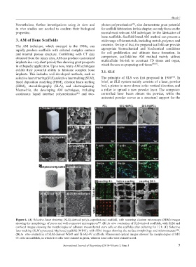
Figure 6. (A) Selective laser sintering (SLS)-derived poly(ε-caprolactone) scaffold, with scanning electron microscope (SEM) images
showing the morphology of pores and well-connected microspheres . (B) In vitro evaluation of SLS-derived scaffolds, with SEM and
[99]
confocal images showing the morphologies of adherent mesenchymal stem cells on the scaffolds after culturing for 12 h. (C) Selective
laser melting (SLM) processed Mg-based scaffold (WE43), with SEM images showing the surface morphology and microstructure [124] .
(D) In vitro evaluation of SLM-derived WE43 and Ti-6Al-4V scaffolds. Fluorescent optical images showed the morphologies of MG
63 cells on scaffolds, in which live cells were stained in green, whereas dead cells were stained in red.
International Journal of Bioprinting (2019)–Volume 5, Issue 1 7