
Page 19 - IJB-5-1
P. 19
Additive manufacturing of bone scaffolds
[7]
to disease transmission and immune rejection . Thus,
idea bone substitutes are urgently demanded for bone
tissue repair in surgical application.
Desired bone substitutes should have a customized external
shape, aiming to avoid the excess removal of bone tissue
at defect sites. More importantly, they also need to have
porous and interconnected pore structure, so as to create
a microenvironment, which is conducive to cell activity
and reproduction [8,9] . To obtain scaffolds with the porous
structure for bone repair, several approaches, such as pore-
forming agent method [10,11] , gas foaming method [12,13] , sol-
gel method [14,15] , and freezing drying method [16,17] , have
been proposed. Although these methods exhibit a certain
ability to fabricate porous structure, they are also with
some limitations, such as inaccurate control of the pore
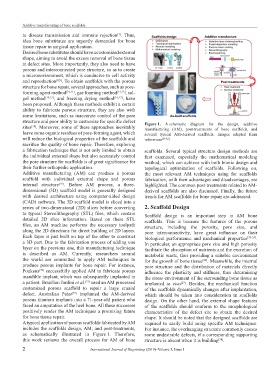
structure and poor ability to customize for specific defect Figure 1. A schematic diagram for the design, additive
sites . Moreover, some of these approaches inevitably manufacturing (AM), post-treatments of bone scaffolds, and
[18]
leave some organic residues of pore-forming agent, which several typical AM-derived scaffolds. Images adapted from
will reduce the biological properties of the scaffolds and references [29-32] .
sacrifice the quality of bone repair. Therefore, exploring
a fabrication technique that is not only limited to obtain scaffolds. Several typical structure design methods are
the individual external shape but also accurately control first examined, especially the mathematical modeling
the pore structure for scaffolds is of great significance for method, which can achieve with both bionic design and
their further orthopedic application. topological optimization of scaffolds. Following on,
Additive manufacturing (AM) can produce a porous the most relevant AM techniques using for scaffolds
scaffold with individual external shape and porous fabrication, with their advantages and disadvantages, are
internal structure . Before AM process, a three- highlighted. The common post-treatments related to AM-
[19]
dimensional (3D) scaffold model is generally designed derived scaffolds are also discussed. Finally, the future
with desired architecture using computer-aided design trends for AM scaffolds for bone repair are addressed.
(CAD) software. The 3D scaffold model is sliced into a
series of two-dimensional (2D) slices before converting 2. Scaffold Design
to typical Stereolithography (STL) files, which contain Scaffold design is an important step in AM bone
detailed 2D slice information. Based on these STL scaffolds. This is because the features of the porous
files, an AM machine performs the necessary toolpath structure, including the porosity, pore size, and
along the 2D directions for direct building of 2D layers. pore interconnectivity, have great influence on their
Each layer is just built on top of the other to construct biological performance and mechanical properties [23-25] .
a 3D part. Due to the fabrication process of adding one In particular, an appropriate pore size and high porosity
layer on the previous one, this manufacturing technique facilitate the absorption of nutrients and the excretion of
is described as AM. Currently, researchers around metabolic waste, thus providing a suitable environment
the world are committed to apply AM techniques to for the growth of bone tissue . Meanwhile, the internal
[26]
produce porous implants for bone repair. For instance, pore structure and the distribution of materials directly
Poukens successfully applied AM to fabricate porous influence the plasticity and stiffness, thus determining
[20]
mandible implant, which was subsequently implanted in the stress environment of the surrounding bone tissue as
a patient. Brazilian Jardini et al. used an AM processed implanted in vivo . Besides, the mechanical function
[21]
[27]
customized porous scaffold to repair a large cranial of the scaffolds dynamically changes after implantation,
defect. Australian Peter implanted the AM-derived which should be taken into consideration in scaffolds
[22]
porous titanium implants into a 71-year-old patient who design. On the other hand, the external shape features
faced an amputation of the heel bone. All these successes of the scaffolds should conform to the morphological
positively render the AM techniques a promising future characteristics of the defect site to obtain the desired
for bone tissue repair. shape. It should be noted that the designed scaffolds are
A typical application of porous scaffolds fabricated by AM required to easily build using specific AM techniques.
includes the scaffolds design, AM, and post-treatments, For instance, the overhanging structure commonly causes
as schematically illustrated in Figure 1. Therefore, some undesirable defects, if a corresponding supporting
this work reviews the overall process for AM of bone structure is absent when it is building .
[28]
2 International Journal of Bioprinting (2019)–Volume 5, Issue 1