
Page 96 - IJB-5-1
P. 96
Gao and Zhou
bulk silver . Prasetyo et al. found that diameters of Ag and aerodynamically assisted bio-jetting and jetted cells
[16]
dots can be reduced on a hydrophobic surface coated exhibited all expected cellular behavior .
[37]
with octadecyltrichlorosilane (OTS), due to lower surface Although electrospray is able to produce relatively
energy. A hydrophobic substrate with OTS coating can smaller droplets, electrospray cannot emit single
also overcome the existence of the coffee-stain effect . microscale droplet on substrate, and sometimes, patterns
[28]
Youn showed that the width of minimum silver line is have broad and non-uniform size distribution. EHD
about 5.8 µm using a tilted nozzle . Wang succeeded high-resolution technique allows to print a controlled
[29]
in printing several passive electrical components, such droplet size and deposit at desired locations. Park et
as coated resistors, interdigitated capacitors, and spiral al. patterned large areas of DNA inks with complex
inductors as shown in Figure 4D-F and the minimum line configuration and feature size with resolutions on
width about 60 µm is achieved using a 110 µm nozzle . the order of 100 nm . Properties of DNA were not
[38]
[30]
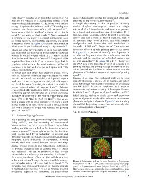
Khalid deposited silver patterns on thick glass substrates adversely affected by this printing process. As shown
and found that the diameter of droplet after the sintering in Figure 5A, a pattern of butterfly was deposited at
process is around 3.6 µm, which is 2.78 times smaller an inherent frequency when the nozzle scan over the
than the internal diameter of nozzle . Kang used EHD substrate and the positions of individual droplets are
[31]
[38]
to print silver lines within 10 µm wide on a large flexible not well controlled . In Figure 5B, a 14 × 14 arrays of
graphene substrate and the sheet resistance of hybrid the DNA dots were deposited by drop-on-demand e-jet
electrode is as low as 4 ohm per unit square with 78% printing method; the driving voltage was turned on and
optical transmittance . off on demand and it offered excellent control over the
[32]
To lower cost and obtain less electromigration effect, placement of individual droplets at expense of printing
colloidal solutions containing copper nanoparticles were speed [38] .
tested . As a result, the resistivity of deposited copper Sutanto et al. used two biological materials to print
[7]
track was 5 times as high as resistivity of bulk copper droplets whose size is about 2 µm on average, and yellow
and the difference of resistivity is attributed to relatively color dots at right bottom corner are formed by overlaying
[11]
porous microstructure of copper track . Rahman two ink dots . It can be considered as a proof to
[7]
et al. applied EHD methods to print a colloidal solution demonstrate registration accuracy of the droplet diameter
[11]
containing copper nanoparticles on a silicon substrate; is within 2 µm . Shigeta et al. used multinozzle EHD
the range of resistivity of the printed copper tracks was inkjet printing system to create micro- and nano-scale
from 5.98 × 10 Ωm to 2.42×10 Ωm. . Han et al. patterns of proteins on flat silica substrates. A structured
−8
[33]
−7
used a nozzle with an inner diameter of 160 µm to print plasmonic analysis is shown in Figure 5C and the study
molten-metal by an EHD method, and a straight metal showed that the printing process does not adversely alter
[39]
line with a diameter of 50 μm was obtained at a plotting the protein structure or function .
speed of 1 mm/s .
[34]
3.2. EHD 3D Printing
3.1.2 Biotechnology Applications
A B
Inkjet printing had been previously employed to process
living cells , but the processing of concentrated
[35]
biosuspensions has been severely limited by cellular
loading and this has resulted in the formation of rather
coarse structures . Jayasinghe et al. for the first time
[36]
used electric field-driven technology to process and
deposit living cells, like Jurkat cell suspension, and human
peripheral blood monocytes in suspension. A strong C
electric field was created between nozzle and ring-
shaped ground electrode and polydisperse distribution
of droplets, indicating that an unstable mode of jetting
was observed. This can be attributed to high electric
conductivity and relatively low viscosity of solution .
[36]
As a result, no adverse effects on either cellular structure
or basic activities of living cells, such as cellular damage Figure 5. Dot diameter is 2 µm (a) butterfly pattern in fluorescence
and irregular rate of division, had been found . To micrographs. (b) 14 × 14 DNA arrays in fluorescence micrograph
[36]
generate a near-mono distribution of cell-bearing droplets printed in drop-on-demand mode (adapted from Park et al. ). (c)
[38]
in stable jetting conditions, Kwok et al. reported a hybrid Fluorescence microscope images of a peacock pattern formed using
processing approach by combining bio-electrosprays different fluorescently proteins (adapted from Shigeta et al. ).
[39]
International Journal of Bioprinting (2019)–Volume 5, Issue 1 7