
Page 79 - IJB-5-2
P. 79
Arab W, et al.
and illness or injury, which influence the balance of 3D bioprinting system [27,28] to fabricate 3D scaffolds
protein synthesis and degradation . Skeletal muscle for the differentiation of myoblast cells. The process of
[6]
is a voluntary moveable tissue that has the ability to 3D bioprinting is believed to enhance the arrangement
convert chemical energy into mechanical energy and then of homogeneous cellular scaffolds and improve cell
transfer it to tendon tissue. It also supports soft tissue proliferation and adhesion for myotube formation. Two
and maintains body posture . In addition, this tissue is sequences of self-assembling peptides are tested and
[7]
responsible for different functions of the body such as analyzed for cell viability, proliferation, and differentiation.
respiration and protection of abdominal viscera, and also The promising results indicate that 3D bioprinting of self-
controls the movement of limbs . Skeletal muscle tissue assembling ultrashort peptides may valuably improve the
[8]
exhibits the native capability to regenerate and repair process of muscle tissue engineering.
through the activation of local satellite cells [8,9] .
However, this ability declines with age as well as in 2. Materials and Methods
clinical conditions such as tumor resection and traumatic Two tetrameric self-assembling peptides CH-01 and
sport injuries including concussions and strains, and CH-02 were custom-synthesized in our Laboratory for
muscular dystrophy that may result in volumetric muscle Nanomedicine. Mouse myoblast cells (C2C12) were
loss (VML). In these injuries, approximately 20% or more obtained from ATCC, USA. The following materials
of the muscle mass is lost [10,11] and, as a result, tissues were ordered from Gibco, USA: Dulbecco’s modified
lose the ability to signal each other and become unable to eagle medium (DMEM), fetal bovine serum (FBS), heat-
repair themselves through natural physiological processes. inactivated horse serum, Dulbecco’s phosphate-buffered
Thus, surgical intervention is needed [12-15] to restore normal saline (PBS) solution, and penicillin-streptomycin
function and prevent the formation of scar tissue , (P/S) antibiotics. An 3-(4,5-Dimethylthiazol-2-yl)-2,5-
[13]
which may lead to muscle atrophy and prevent muscle diphenyltetrazolium bromide (MTT) cell proliferation
regeneration . Around the world, millions of people are assay kit and a LIVE/DEAD Viability/Cytotoxicity kit
[16]
affected by these clinical conditions which cause significant were purchased from Promega, USA. Immunostaining
social and economic problems [17,18] . As such, alternative antibody myosin heavy chain (MHC) was purchased
technologies are urgently needed for the reconstruction of from Abcam. Cell culture flasks and 96-well plates were
skeletal muscle tissues that have experienced VML and ordered from Corning, USA.
need to regenerate new functional tissue [10,19] .
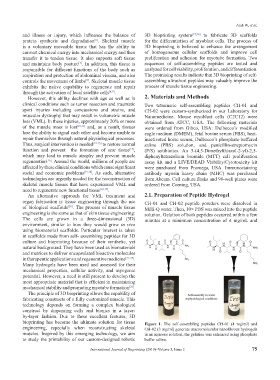
An alternative approach for VML treatment and 2.1. Preparation of Peptide Hydrogel
organ fabrication is tissue engineering through the use CH-01 and CH-02 peptide powders were dissolved in
of biological scaffolds . The process of muscle tissue Milli-Q water. Then, 10× PBS was mixed into the peptide
[20]
engineering is the same as that of skin tissue engineering: solution. Gelation of both peptides occurred within a few
The cells are grown in a three-dimensional (3D) minutes at a minimum concentration of 4 mg/mL and
environment, similar to how they would grow in vivo
using biomaterial scaffolds. Particular interest is taken
in scaffolds made from self- assembling peptides for 3D
culture and bioprinting because of their synthetic, yet
natural background. They have been used as biomaterials
and matrices to deliver encapsulated bioactive molecules
in therapeutic applications and regenerative medicine [21-25] .
Many hydrogels have been used and assessed for their
mechanical properties, cellular activity, and myogenic
potential. However, a need is still present to develop the
most appropriate material that is efficient in maintaining
mechanical stability and promoting myotube formation .
[26]
The principle of 3D bioprinting allows the capability of
fabricating constructs of a fully customized muscle. This
technology depends on forming a complex biological
construct by dispensing cells and bionics in a layer-
by-layer fashion. Due to these excellent features, 3D
bioprinting has become the ultimate solution for tissue Figure 1. The self-assembling peptides CH-01 (4 mg/ml) and
engineering, especially when reconstructing skeletal CH-02 (3 mg/ml) generate macromolecular nanofibrous hydrogels
muscles. Inspired by this emerging technology, we aim in an aqueous solution, the gelation was enhanced using phosphate
to study the printability of our custom-designed robotic buffer saline.
International Journal of Bioprinting (2019)–Volume 5, Issue 2 75