
Page 105 - IJB-6-3
P. 105
Bozo, et al.
0.002, respectively. Materials resorbed form implantation. Since day 15, a highly vascularized
their surfaces and within a zone of the direct connective tissue capsule with a thickness of
contact to soft tissues. The diameter and volume 50 – 70 μm formed around implants. However,
gradually reduced and the “surface-to-volume” cellular and tissue elements did not penetrate into
ratio increased both in 3D printed scaffolds and OCP structure. Starting from day 45, in some parts
gene-activated implants. An average diameter of the implants there were defects in the structure,
decreased more intensively with an abrupt drop their number and size gradually increased by day
by day 40 and a subsequent stabilization and a 180 by forming “cavities of dissolution” with
smooth decline in the test group, while its change a diameter of 30 – 50 μm and scalloped edges
being almost linear in the control group. There that indicated biodegradation. There were no
was a two-fold volume reduction by day 60 in multinucleated foreign bony giant cells. On day
both groups (Figure 4). 180 after implantation, connective tissue grew
We found no histological signs of inflammation inside the implants to 50 – 70 μm from a surface
in the area of 3D printed OCP (control) (Figure 4).
A
B
C D E
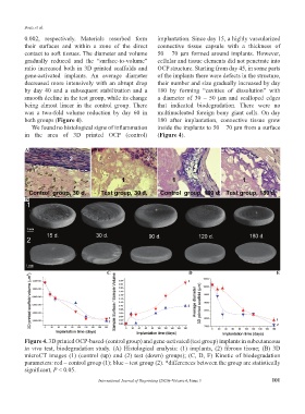
Figure 4. 3D printed OCP-based-(control group) and gene-activated (test group) implants in subcutaneous
in vivo test, biodegradation study. (A) Histological analysis: (1) implants, (2) fibrous tissue; (B) 3D
microCT images (1) (control (up) and (2) test (down) groups); (C, D, F) Kinetic of biodegradation
parameters: red – control group (1); blue – test group (2). *differences between the group are statistically
significant, P < 0.05.
International Journal of Bioprinting (2020)–Volume 6, Issue 3 101