
Page 106 - IJB-6-3
P. 106
3D printed gene-activated implants for bone regeneration
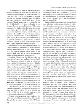
The biodegradation study of personalized gene- scaffolds defused without a border between newly
activated constructs demonstrated implants structure formed bone tissue and the implant. Calcium to
disintegration and connective tissue growing into phosphorus ratio (Ca/P) in the areas of 3D printed
them from day 15 after implantation. A capsule implants and newly formed bone tissue was 2.06
covering the implants included active fibroblasts and 1.83 and 1.90 and 2.23 in 3 and 6 months after
and was intensively vascularized with a larger surgery, respectively.
number of blood vessels than in the control group Based on histological analysis, a gene-activated
(P = 0.021). In 45 – 120 days, we found a further bone substitute surface contacted directly with a
structure disintegration and thinning of 3D printed newly formed woven bone tissue without forming a
gene-activated implant edges. Later on, all implants connective tissue capsule in 3 months after surgery.
were highly porous: optically empty vacuoles There were both macropores corresponding to
occupied a significant volume (about 60%), their prefabricated canals and numerous micropores
diameter achieved 200 – 250 μm. Connective tissue in the implant structure. Trabeculae of a
in-grew to a depth of 100 – 150 μm (Figure 4). As newly formed bone tissue enlaced the implant,
in the control, there were no signs of inflammation. directly extending into macro- and micropores.
In general, personalized gene-activated materials Vascularized bone tissue had been growing from
possessed a profile of more evident bioresorption both periosteum and endosteum sides. Bone rods
and a slightly higher rate of biodegradation. spread from the implant to a diaphysis wall in the
None of animals died during the experiment until form of bridges (Figure 5). Bone trabeculae arose
a planned sacrifice. Wounds healed in three animals directly from the implant both in the periphery and
without abnormalities in 10 days after surgery. in its depth; newly formed trabeculae adhered to
Since that time the animals started resting on the a rarefied implant surface, their side contacting
operated limb; one animal had a post-operative with the material had irregular edge, an opposite
wound infection after tibia reconstruction. side was characterized by a smooth surface with
Based on CT findings, the tibia integrity was osteoblasts and bone lining cells involved. Fusing
restored in all cases, and implants integrated with trabeculae of newly formed bone tissue constituted
bone fragments without forming a connective a mesh structure, neither evidence of woven bone
tissue capsule. Bone thickness increased within the remodeling into lamellar tissue, nor osteoclastic
area of intervention due to a pronounced periosteal resorbtions occurred at this time point. A fibrous
callus; its diameter achieved 31.5 and 40.3 mm tissue was detected only within inter-trabecular
in the greatest dimension in 3 and 6 months after spaces. A newly formed diaphysis wall consisted
surgery, respectively, with an initial diameter of of a spongy bone with bone marrow elements in
being <20 mm. The implants retained their initial inter-trabecular spaces. There was a pronounced
shape with structure becoming heteromorphic. In periosteal response with woven bone tissue
addition to canals filled with newly formed tissues trabeculae formation within basal regions of the
of bone density, we detected some cracks without periosteum.
fragment disintegration in the implant structure In 6 months after surgery, personalized gene-
(Figure 5). The implants length reduced to 26 and activated bone substitutes were completely
24 mm, whereas their diameter to 15 and 14 mm integrated into the tibia proximal and distal
in 3 and 6 months after surgery, respectively. An fragments and significantly rarefied around initial
average tissue density within a tibia reconstruction perforations. Macro- and micropores as well as
zone was 1362 ± 617.6 HU in 3 months and a peripheral implant surface were covered with
1332 ± 572.2 HU in 6 months, with the initial trabeculae of a newly formed bone tissue. By
implant density of more than 2000 HU. this time point, the bone tissue directly in implant
Optimal osteointegration was confirmed by perforations as well as along its perimeter remodeled
SEM and a histologic examination. SEM results into lamellar forming osteons even in the implant
showed that a crystal structure of OCP-based macropores. There were no cells responsible for
102 International Journal of Bioprinting (2020)–Volume 6, Issue 3