
Page 110 - IJB-6-3
P. 110
3D printed gene-activated implants for bone regeneration
First, in our work, we have realized a The next task of the present work involved the
combination and further development of the production of gene-activated implants based on
processes involving chemical interaction between the OCP and plasmid DNA with VEGFА gene.
TCP agglomerated particles and “ink” based on According to fluorimetry data, the concentration
diluted phosphoric acid, followed by chemical of plasmid DNA bound to 3D printed scaffolds
post-treatment of the printed DCPD structure at was 52.74 ± 1.76 ng/mg that correlated with our
physiological temperatures . DCPD structure previous findings related with OCP granule-based
[11]
[32]
can be further transformed into OCP phase. bone substitute .
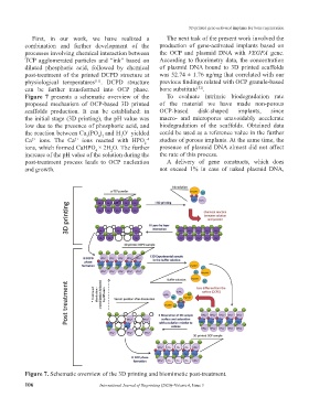
Figure 7 presents a schematic overview of the To evaluate intrinsic biodegradation rate
proposed mechanism of OCP-based 3D printed of the material we have made non-porous
scaffolds production. It can be established: in OCP-based disk-shaped implants, since
the initial stage (3D printing), the pH value was macro- and micropores unavoidably accelerate
low due to the presence of phosphoric acid, and biodegradation of the scaffolds. Obtained data
the reaction between Ca (PO ) and H O yielded could be used as a reference value in the further
+
4 2
3
3
Ca ions. The Ca ions reacted with HPO studies of porous implants. At the same time, the
-4
2+
2+
2
ions, which formed CaHPO × 2H O. The further presence of plasmid DNA almost did not affect
2
4
increase of the pH value of the solution during the the rate of this process.
post-treatment process leads to OCP nucleation A delivery of gene constructs, which does
and growth. not exceed 1% in case of naked plasmid DNA,
Figure 7. Schematic overview of the 3D printing and biomimetic post-treatment.
106 International Journal of Bioprinting (2020)–Volume 6, Issue 3