
Page 108 - IJB-6-3
P. 108
3D printed gene-activated implants for bone regeneration
the mandible in 3 months after surgery; irregular side. There were evident signs of periosteal
implant surface was directly surrounded by a osteoblasts were nearby the zone of implantation
newly formed bone tissue of a mixed structure (Figure 6). A richly vascularized connective tissue
(woven and lamellar) without a connective was detected in inter-trabecular spaces. No cellular
tissue capsule. There was a typical tendency for or tissue structures were identified in the implant
osteon-like structures and circular trabeculae to central zones. There were single osteoclasts on
be formed. There were no cells on the surface of a newly formed bone trabeculae surface on the
bone trabeculae in contact with the bone substitute, side of connective tissue. A similar picture was
while active osteoblasts detected on the opposite observed in 3D printed OCP implants without gene
A
B
C
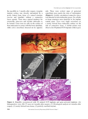
Figure 6. Mandible reconstructed with 3D printed OCP implants and gene-activated implants. (A)
Intraoperation view; (B) CT scans in 6 months after surgery; (C) Histological analysis in 6 months after
surgery: (1) newly formed bone tissue; (2) partially resorbed implant.
104 International Journal of Bioprinting (2020)–Volume 6, Issue 3