
Page 109 - IJB-6-3
P. 109
Bozo, et al.
constructs; however, periosteal osteogenesis was developed, probably due to the attractive idea to
less evident. create a fully functional tissue or organ ex vivo
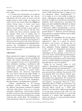
In 6 months after implantation, the peripheral for subsequent clinical transplantation [6,10,16,17]
part of gene-activated materials was actively or for usage as an in vitro model to study the
[18]
substituted with bone tissue. A border with the disease pathogenesis and drugs development .
implant along its entire length was composed of However, in addition to well-known technological
newly formed bone tissue trabeculae (Figure 6) problems, tissue engineering is associated with
without fibrous tissue. A woven bone tissue high costs and considerable difficulties in clinical
[9]
remodeled into lamellar tissue. The implant translation from a regulatory point of view .
surface was rarefied and had a great number of Furthermore, additive manufacturing is actively
optically empty cavities sized to 300 μm. Still, used to produce personalized bone implants with
no cellular and tissue elements extended into growth factors [19,20] . However, protein molecules,
the material. There were no cellular signs of being short-lived, and short-distant, cannot exhibit
osteoclastic resorption. Inter-trabecular spaces their full biological effect.
were in part colonized with hematopoietic bone Although 3D bioprinting has become more
marrow. There was an optimal osteointegration in complex in an attempt to combine different
[21]
3D printed OCP implants with similar dynamics approaches , there is a search for simpler
of bone tissue rearrangement into lamellar tissue; alternatives that involves, for instance, the use
however, the colonization of inter-trabecular of gene constructs for creating personalized
spaces with blood-forming (hematopoietic) bone bone substitutes. Despite significant advances
marrow was less active. in the research of standardized gene-activated
matrices [22-24] and additive manufacturing
4 Discussion technologies for bone grafting, to date there are
only a few studies related with personalized gene-
Bone reconstructive surgery in traumatology and activated tissue substitutes [25,26] , and none of them
orthopedics, neurosurgery, oral and maxillofacial described calcium phosphate-based ceramic being
surgery still remains challenging. Small defects used as a scaffold without any hydrogels or other
can be managed with the use of osteoconductive materials that are easy to be printed.
bone substitutes, including a combination with Based on our previous results in OCP
“improvised” techniques for biologic activity studies , 3D printing of OCP-based implants
[11]
[27]
enhancement such as mixing with autologous and standardized gene-activated materials [23,28] , we
bone fragments, platelet-, or growth factor- have started the development of a personalized
enriched plasma, etc. However, large/critical gene-activated bone substitute based on the OCP
size bone defects and alveolar ridge atrophy of and plasmid DNA that delivers VEGFA gene,
significant size are characterized by “osteogenic applicable for large bone defects substitution
insufficiency,” a loss of cambial cellular elements and guided bone regeneration. We expected the
and/or factors, invloved in bone regeneration, increased level of VEGFA to promote angiogenesis
therefore intensive osteoinduction is required . and reparative osteogenesis. Moreover, a direct
[9]
Additive manufacturing or 3D printing stimulating effects of VEGF on proliferation and
techniques provide substantial opportunities differentiation of bone cells and non-canonic
[29]
for effective personalized treatment of patients intracrine effects specific for a VEGF gene
with large bone defects. However, to overcome transfer were described. Additional prerequisites
[30]
osteogenic insufficiency, custom-made implants to use this gene construct in the study were our
should be combined with osteogenesis-stimulating previously obtained clinical data on a successful
factors such as living cells, growth factors, or gene treatment of a patient with mandibular non-
constructs that encode them. 3D bioprinting of unions with the use of a gene-activated material,
tissue-engineered constructs are most intensively delivering VEGFA .
[28]
International Journal of Bioprinting (2020)–Volume 6, Issue 3 105