
Page 314 - IJB-10-6
P. 314
International Journal of Bioprinting Bioprinted plasma biocarriers for MSC delivery
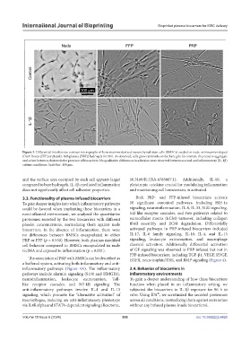
Figure 3. Differential interference contrast micrographs of bone marrow-derived mesenchymal stem cells (BMSCs) seeded on nude or biocarrier-doped
(fresh frozen [FFP] or platelet-rich plasma [PRP]) hydrogels for 96 h. As observed, cells grow randomly on the bare gels. In contrast, they tend to aggregate
and orient between clusters in the presence of biocarriers. No qualitative differences in adhesion were observed between normal and inflammatory (IL-1β)
culture conditions. Scale bar: 100 µm.
and the surface area occupied by each cell appears larger 10.3180/R-HSA-6785807.1). Additionally, IL-10, a
compared to bare hydrogels. IL-1β-mediated inflammation pleiotropic cytokine crucial for modulating inflammation
does not significantly affect cell adhesion properties. and maintaining cell homeostasis, is activated.
3.3. Functionality of plasma-infused biocarriers Both PRP- and FFP-infused biocarriers activate
To gain deeper insights into which inflammatory pathways 10 significant canonical pathways, including HIF-1a
could be favored when implanting these biocarriers in a signaling, neuroinflammation, IL-8, IL-33, S100 signaling,
non-inflamed environment, we analyzed the quantitative toll-like receptor cascades, and two pathways related to
proteomes secreted by the two biocarriers with different extracellular matrix (ECM) turnover, including collagen
platelet concentrations, normalizing them against nude fibril assembly and ECM degradation. Differentially
biocarriers. In the absence of inflammation, there were activated pathways in PRP-infused biocarriers included
no differences between BMSCs encapsulated in either IL-17, IL-6 family signaling, IL-10, IL-4, and IL-13
PRP or FFP (p = 0.550). However, both plasmas modified signaling, leukocyte extravasation, and macrophage
cell behavior compared to BMSCs encapsulated in nude classical activation. Additionally, differential activation
GelMA and exposed to inflammation (p = 0.001). of GF signaling was observed in PRP-infused but not in
FFP-infused biocarriers, including TGF-β1, VEGF, PDGF,
The association of PRP with BMSCs can be described as EGFR, neurotrophin/TRK, and BMP signaling (Figure 4).
a buffered system, activating both inflammatory and anti-
inflammatory pathways (Figure 4A). The inflammatory 3.4. Behavior of biocarriers in
pathways include alarmin signaling (S100 and HMGB1), inflammatory environments
neuroinflammation, leukocyte extravasation, Toll- To gain a deeper understanding of how these biocarriers
like receptor cascades, and NF-kB signaling. The function when placed in an inflammatory setting, we
anti-inflammatory pathways involve IL-4 and IL-13 subjected the biocarriers to IL-1β exposure for 96 h in
®
signaling, which promote the “alternative activation” of vitro. Using IPA , we scrutinized the secreted proteomes
macrophages, inducing an anti-inflammatory phenotype across all conditions, normalizing them against secretomes
via IL4R alpha and STAT6-dependent signaling (Reactome, without any infused plasma (nude biocarriers).
Volume 10 Issue 6 (2024) 306 doi: 10.36922/ijb.4426