
Page 316 - IJB-10-6
P. 316
International Journal of Bioprinting Bioprinted plasma biocarriers for MSC delivery
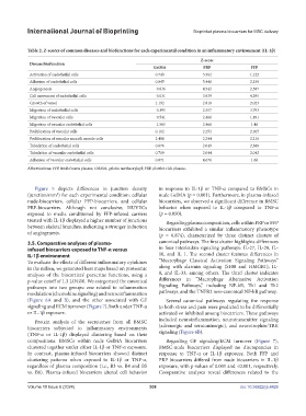
Table 2. Z-scores of common diseases and biofunctions for each experimental condition in an inflammatory environment (IL-1β)
Z-score
Disease/biofunction
GelMA PRP FFP
Activation of endothelial cells 0.749 5.562 1.122
Adhesion of endothelial cells 0.045 5.446 2.339
Angiogenesis −0.836 4.542 2.587
Cell movement of endothelial cells −0.031 3.939 4.295
Growth of vessel 1.192 2.818 2.023
Migration of endothelial cells −0.395 2.557 3.793
Migration of vascular cells −0.541 2.466 1.491
Migration of vascular endothelial cells −1.505 2.466 1.46
Proliferation of vascular cells 0.102 2.252 2.105
Proliferation of vascular smooth muscle cells 2.488 2.244 2.216
Tubulation of endothelial cells 0.676 2.049 2.389
Tubulation of vascular endothelial cells 0.719 2.044 2.042
Adhesion of vascular endothelial cells 0.071 4.676 1.66
Abbreviations: FFP, fresh frozen plasma; GelMA, gelatin methacryloyl; PRP, platelet-rich plasma.
Figure 5 depicts differences in junction density in response to IL-1β or TNF-α compared to BMSCs in
(junction/mm ) for each experimental condition: cellular nude GelMA (p = 0.001). Furthermore, in plasma-infused
2
nude-biocarriers, cellular FFP-biocarriers, and cellular biocarriers, we observed a significant difference in BMSC
PRP-biocarriers. Although not conclusive, HUVECs behavior when exposed to IL-1β compared to TNF-α
exposed to media conditioned by FFP-infused carriers (p = 0.050).
treated with IL-1β displayed a higher number of junctions Regarding plasma composition, cells within PRP or FFP
between skeletal branches, indicating a stronger induction biocarriers exhibited a similar inflammatory phenotype
of angiogenesis. (p = 0.874), characterized by three distinct clusters of
3.5. Comparative analyses of plasma- canonical pathways. The first cluster highlights differences
infused biocarriers exposed to TNF-α versus in four interleukin signaling pathways: IL-27, IL-20, IL-
IL-1β environment 10, and IL 1. The second cluster features differences in
To evaluate the effects of different inflammatory cytokines “Macrophage Classical Activation Signaling Pathways”
in the milieu, we generated heat maps based on proteomic along with alarmin signaling (S100 and HMGB1), IL-
analyses of the biocarriers’ paracrine functions, using a 8, and IL-33, among others. The third cluster indicates
p-value cutoff of 1.3 LOG10. We categorized the canonical differences in “Macrophage Alternative Activation
pathways into two groups: one related to inflammation Signaling Pathways,” including NF-kB, Th1 and Th2
modulation (chemokine signaling) and neuroinflammation pathways, and the TNFR2 non-canonical NF-kB pathway.
(Figure 6A and B), and the other associated with GF Several canonical pathways regulating the response
signaling and ECM turnover (Figure 7), both under TNF-α to both stress and pain were predicted to be differentially
or IL-1β exposure. activated or inhibited among biocarriers. These pathways
Protein analysis of the secretomes from all BMSC included neuroinflammation, neurotransmitter signaling
biocarriers subjected to inflammatory environments (adrenergic and serotoninergic), and neurotrophin/TRK
(TNF-α or IL-1β) displayed clustering based on their signaling (Figure 6B).
compositions. BMSCs within nude GelMA biocarriers Regarding GF signaling/ECM turnover (Figure 7),
clustered together under either IL-1β or TNF-α exposure. BMSC-nude biocarriers displayed no discrepancies in
In contrast, plasma-infused biocarriers showed distinct response to TNF-α or IL-1β exposure. Both FFP and
clustering patterns when exposed to IL-1β or TNF-α, PRP biocarriers differed from nude biocarriers in IL-1β
regardless of plasma composition (i.e., B3 vs. B4 and B5 exposure, with p-values of 0.001 and <0.001, respectively.
vs. B6). Plasma-infused biocarriers altered cell behavior Comparative analyses reveal differences related to the
Volume 10 Issue 6 (2024) 308 doi: 10.36922/ijb.4426