
Page 481 - IJB-10-6
P. 481
International Journal of Bioprinting Nanomaterial-bioinks for DLP bioprinting
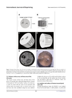
Figure 1. Design and dimension of the 3D model. (A and B) A cylindrical 3D model with interconnective pores (pore diameter: 500 μm) arranged in a
regular grid and larger pores (pore diameter: 2.5 mm): top view (A) and front view (B). (C and D) The 3D model from different perspectives: Figure 1C
(perspective A) and Figure 1D (perspective B). (E) Light microscopy image of a printed GelMaBB construct. (F) Image of the printed GelMaBB construct.
Scale bars: 3.5 cm (C–F). Magnification: 4×. Abbreviations: GelMa: Gelatin methacrylamide; BB: Brilliant Black.
2.2. Volume, surface area, and base area of the Additionally, the porous 3D model was sliced into sections
3D model at 100 µm intervals, and the surface area of each section
The overall volume, surface area, and base area of the was calculated to determine the minimum, mean, and
porous 3D model were calculated using Blender (version maximum areas of the sections.
2.93.4) with the “3D Print Toolbox” add-on. For reference, 2.3. Isolation and culture of human mesenchymal
a solid 3D model with the same overall geometry but stem cells
without pores was created, and its volume, surface area, Human mesenchymal stem cells (hMSCs) were isolated
and base area were calculated using the same method. from the femoral heads of adult patients, as previously
Volume 10 Issue 6 (2024) 473 doi: 10.36922/ijb.4015