
Page 482 - IJB-10-6
P. 482
International Journal of Bioprinting Nanomaterial-bioinks for DLP bioprinting
described. 67,68 The use of human tissue was approved by nanoparticles (particle size: < 150 nm) (Sigma-Aldrich,
the local ethical advisory board of the University Medical USA) were added to the bioink at a concentration of 50 mg/
Center in Kiel (approval number: D459/13) and included mL. Before printing, the resulting ink (GelMaBB-CaP) was
the consent of the individual donors. Cells were precultured incubated at 37°C for 2 h to allow swelling of CaP particles
in osteogenic differentiation medium (ODM), i.e., DMEM and a homogeneous consistency of the bioink. In addition,
F12 (PAN Biotech, Germany), supplemented with 10% the GelMa bioink was modified with GO, provided by the
fetal bovine serum (FBS; PAN Biotech, Germany), 1% technical faculty at Kiel University. GO was added at a final
penicillin/streptomycin (Biochrom, Germany), 0.1 µM concentration of 0.5 mg/mL to the bioink (GelMaGO). The
dexamethasone (Sigma-Aldrich, United States of America bioinks were loaded with 3 × 10 hMSC/mL bioink before
6
[USA]), 50 µM ascorbic acid (Sigma-Aldrich, USA), and the DLP bioprinting process.
10 µM ß-glycerolphosphate (Sigma-Aldrich, USA), to
induce osteogenic differentiation. For the printing process, 2.5. Bioprinting of constructs using DLP technology
hMSCs (passages 3 and 4) were added to different types Tissue constructs were printed by DLP using LumenX
of bioinks. (Cellink, Sweden) equipped with LightField software. To
print under sterile conditions, the Lumen X printer was
2.4. Formulation and modifications of placed under a laminar flow bench, and sterile equipment
different bioinks was used. The printbed and printhead were further treated
Different types of GelMa-based bioinks were formulated with 70% ethanol. For the printing process, 1 mL bioink
and compared in terms of their printability and was loaded into the printer, resulting in three constructs
biocompatibility, both before and after modification with for the given design. The printbed temperature was set to
nanomaterials. In this context, a commercially available 37°C. Details of the printing parameters are listed in Table
GelMa bioink (GelMa PhotoInk; Cellink, Sweden) served S2, Supporting Information and were identical for all
as reference material for the evaluation process. The basis bioinks. The 3D model was sliced into layers of 100 µm
of the different bioinks was formulated using sterile GelMa height. The first layer was exposed with a factor of 4. The
with a methacrylation degree of 80% (GelMa; Gelomics, exposure time of the light was 9 s, with an intensity of 70%
Australia). In brief, GelMa was dissolved in ODM at a (33.6 mW/cm ). After the printing process, gel-based tissue
2
concentration of 100 mg/mL. Lithium-phenyl-2,4,6- constructs were transferred to a 24-well plate (Sarstedt,
tri-methylbenzoylphosphinate (LAP; Sigma-Aldrich,
USA) was used in all bioinks as a photo-initiator at a Germany) and washed three times with phosphate-
final concentration of 5 mg/mL. In contrast, the photo- buffered saline (PBS) (Gibco by Life Technologies, USA)
69
absorber concentration varied between the different for 10 min each. Each construct was then placed in 2 mL
bioinks (Table 1). As indicated in Table 1, Brilliant Black of ODM and further cultured at 37°C and 5% CO 2 for
(BB) (Sigma-Aldrich, USA) was used as a photo-absorber the indicated time points. For the controls, samples with
at a final concentration of 0.5 mg/mL, dissolved in and without cells were printed as indicated in the sections
ODM. The components of the bioink (LAP and BB) were for the individual analyzing methods. Scaffolds with or
individually filtered (Filtropur 0.2 µm; Sarstedt, Germany) without cells were treated and cultured in the same way for
for sterilization. a better comparison.
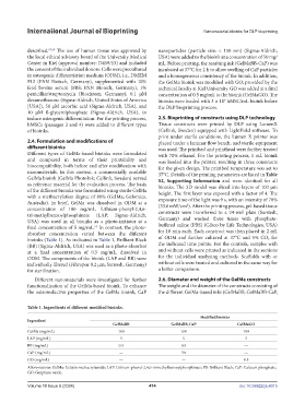
Different nanomaterials were investigated for further 2.6. Diameter and weight of the GelMa constructs
functionalization of the GelMa-based bioink. To enhance The weight and the diameter of the constructs consisting of
the osteoinductive properties of the GelMa bioink, CaP the different GelMa-based inks (GelMaBB, GelMaBB-CaP,
Table 1. Ingredients of different modified bioinks.
Modified bioinks
Ingredient
GelMaBB GelMaBB-CaP GelMaGO
GelMa (mg/mL) 100 100 100
LAP (mg/mL) 5 5 5
BB (mg/mL) 0.5 0.5 —
CaP (mg/mL) — 50 —
GO (mg/mL) — — 0.5
Abbreviations: GelMa: Gelatin methacrylamide; LAP: Lithium-phenyl-2,4,6-trimethylbenzoylphosphinate; BB: Brilliant Black; CaP: Calcium phosphate;
GO: Graphene oxide.
Volume 10 Issue 6 (2024) 474 doi: 10.36922/ijb.4015