
Page 487 - IJB-10-6
P. 487
International Journal of Bioprinting Nanomaterial-bioinks for DLP bioprinting
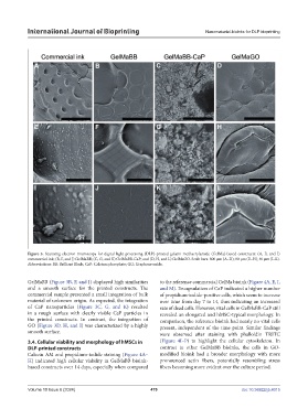
Figure 3. Scanning electron microscopy for digital light processing (DLP)-printed gelatin methacrylamide (GelMa)-based constructs: (A, E, and I)
commercial ink; (B, F, and J) GelMaBB; (C, G, and K) GelMaBB-CaP; and (D, H, and L) GelMaGO. Scale bars: 800 µm (A–D); 80 µm (E–H); 40 µm (I–L).
Abbreviations: BB: Brilliant Black; CaP: Calcium phosphate; GO: Graphene oxide.
GelMaBB (Figure 3B, F, and J) displayed high similarities to the reference commercial GelMa bioink (Figure 4A, E, I,
and a smooth surface for the printed constructs. The and M). Encapsulation of CaP indicated a higher number
commercial sample presented a small integration of bulk of propidium-iodide-positive cells, which seem to increase
material of unknown origin. As expected, the integration over time from day 7 to 14, thus indicating an increased
of CaP nanoparticles (Figure 3C, G, and K) resulted rate of dead cells. However, vital cells in GelMaBB-CaP still
in a rough surface with clearly visible CaP particles in revealed an elongated and hMSC-typical morphology. In
the printed constructs. In contrast, the integration of comparison, the reference bioink had nearly no vital cells
GO (Figure 3D, H, and I) was characterized by a highly present, independent of the time point. Similar findings
smooth surface. were observed after staining with phalloidin TRITC
3.4. Cellular viability and morphology of hMSCs in (Figure 4I–P) to highlight the cellular cytoskeleton. In
DLP-printed constructs contrast to other GelMaBB bioinks, the cells in GO-
Calcein AM and propidium-iodide staining (Figure 4A– modified bioink had a broader morphology with more
H) indicated high cellular viability in GelMaBB bioink- pronounced actin fibers, potentially resembling stress
based constructs over 14 days, especially when compared fibers becoming more evident over the culture period.
Volume 10 Issue 6 (2024) 479 doi: 10.36922/ijb.4015