
Page 153 - IJB-7-3
P. 153
Zhou, et al.
A B
C
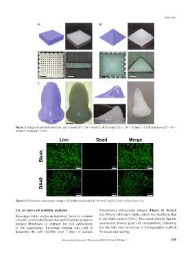
Figure 5. Images of printed constructs. (A) Cuboid (20 × 20 × 4 mm ). (B) Pyramid (20 × 20 × 10 mm ). (C) Human nose (28 × 18 ×
3
3
10 mm ). Scale bars: 5 mm.
3
Figure 6. Fluorescent microscopic images of fibroblast cultured with DA40 (5 mg/mL) and with medium only.
3.6. In vitro cell viability analysis Fluorescence microscopy images (Figure 6) showed
Biocompatibility is also an important factor to evaluate that 90% of cells were viable, which was similar to that
a bioink; a cell viability test was performed using human in the blank control (92%). This result showed that the
primary fibroblasts to estimate the cell cytotoxicity copolymers possess good cell compatibility, indicating
of the copolymers. Live/dead staining was used to that the inks may be printed to biodegradable scaffold
determine the cell viability over 3 days of culture. for tissue engineering.
International Journal of Bioprinting (2021)–Volume 7, Issue 3 149