
Page 27 - IJB-7-3
P. 27
Ren, et al
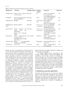
Table 2. Performance comparison of different bioinks for organoids
Bioink types Cell types Printing methods Gelation Properties References
method
Collagen-based Human stem cell–derived Extrusion pH Better biocompatibility [33]
cardiomyocytes Slow gelation rate
Low mechanical properties
ECM-based Human induced pluripotent Extrusion Light Better biocompatibility [36]
stem cells (hiPSCs) Better functionality
Alginate-based HepaRG Extrusion Temperature/ Easy to prepare [52]
ion Fast gelation
Better cytocompatibility
Hyaluronic acid- Primary cell liver Extrusion Che mic al Better biocompatibility [28]
based crosslinking Slower gelation lower
Mechanical properties
Agarose-based Human umbilical vein Extrusion Che mic al Good gel forming ability [25]
smooth muscle cells crosslinking Good mechanical properties
(HUVSMCs) and biological tolerance
Human skin fibroblasts Limited ability to support
(HSFs) cell growth
Fibrin-based Human adipose-derived Laser-assisted Thrombin Better biocompatibility, [30]
stem cells (ASCs) biodegradability
Endothelial colony-forming Poor mechanical properties
cells (ECFCs)
Cellulose-based Human nasoseptal Inkjet Temperature Environmentally sensitive [29]
chondrocytes cells (hNCs) Easy to gel
Gelatin-based HepG2 cells Extrusion Light Better biodegradability [31]
and remodeling
alginate ink alone. The application of bioink with two protein obtained by the partial hydrolysis of collagen and
or even three biomaterials will improve the stability of is homologous with collagen.
polymer systems, tissues, and organoid constructs and will Its strength depends on the concentration of the
be more beneficial for cell proliferation, differentiation, solution. Gelatin exhibits sufficient degradability and
and self-organization. Hyaluronic acid (HA) is a natural remodeling. ECM bioink, formed by crushing the
ECM. HA gels slowly, have low mechanical properties removed cellular tissue, dissolving it in buffer, and adding
after gel formation, and are usually double cross-linked other easy-to-form gels, is the most suitable bioink for
or chemically modified to improve its mechanical cell survival. Matrigel™, an ECM secreted from murine
properties. Skardal et al. developed a versatile HA and Engelbreth–Holm–Swarm tumors, is the most commonly
gelatin-based hydrogel system to print primary liver used ECM for bioprinting. Salvador et al. used hydrogels
spheroids . Carboxymethylcellulose (CMC) is a semi- composed of alginate, gelatin, and matrix gel-controlled
[28]
flexible polysaccharide derived from cellulose. CMC can fractions for bioprinting tumor models to maintain and
be converted into environmentally sensitive hydrogels prolong patient-derived tumor spheres in culture without
[31]
by changing its concentrations and molecular weights, disrupting tumor sphere formation .
as appropriate. Markstedt et al. combined nanofibrillated 4. Bioprinting organoids applications
cellulose–alginate complexes and chondrocytes to prepare
ear-shaped and curved-moon scaffolds . Fibrin is a pro- Organoids and bioprinting are two of the most popular
[29]
coagulant protein. It is enzymatically thrombinized to areas of tissue engineering. Although the use of bio-3D
prepare hydrogels with adequate biocompatibility and printers to print organoids is nascent, the combination of
biodegradability. Gruene et al. used laser-assisted bioprinting and organoids has demonstrated successful
[30]
bioprinting to produce stable vascular networks using examples, indicating their promising future. Here, we
natural hydrogels composed of fibrin precursors and HA present the current state of research on bioprinting of
as cell carriers and environmental materials. Gelatin is a organoids.
International Journal of Bioprinting (2021)–Volume 7, Issue 3 23