
Page 29 - IJB-7-3
P. 29
Ren, et al
researchers at the University of Minnesota in the U.S. using a single protocol. Kidney unit patterns and cell ratios
have recently 3D printed the first ever centimeter-sized may also fluctuate between experiments. In the field of
heart organoids. They optimized a specialized bioink, bioprinting, the construction of kidney organoids likely
made from ECM proteins and human stem cells, to print yields satisfactory results. Jennifer et al. used bioprinting
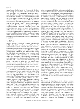
into ventricular structures. The corresponding stem cells to construct a functional 3D kidney structure containing
were first expanded to high cell density on the ventricular living human epithelial cells that form the surface of
structures. Then, the cells were differentiated into the renal tubules (Figure 3A, B and C). Organovo
[43]
cardiomyocytes, with critical cell density and the ability to recently developed a proximal tubule-like tissue that
make the cells beat like a heart . This is a major advance was bioprinted as a layered structure in a well membrane
[39]
in organoid studies of the heart to bioprint stem cells in by mixing fibroblasts and HUVECs with a proprietary
a tissue synergistic manner and to be able to direct their heat-responsive hydrogel. After 3 days of culture, renal
differentiation into cardiomyocytes in similar situations PTECs were inoculated onto the bioimprinted layer. On
with in vivo stem cells adjacent to each other. While their maturation, the kidney cells exhibited a microvascular
printed cardiac muscle models demonstrated encouraging network with tight junctions and cell polarization
results in small kinetic models, this is insufficient in large (Figure 3D, E, and F). In nephrotoxicity tests of mature
animal models with thicker myocardial walls and more tissues, the metabolism of renal cells and cellular
demanding vascularization; therefore, further exploration activity produced greater adverse effects with increasing
is required. concentrations of cisplatin . Bioprinting facilitates the
[44]
precise control of cell deposition in a 3D space in terms
4.2. Kidney of the speed and scale, which could lead to a significant
Kidney organoids primarily comprise metanephros reduction in variability between batches of constructed
(MM) cells, which have been successfully used for kidney organoids and even a breakthrough in scale
nephron-related disease modeling and drug screening. from millimeters to centimeters. Recently, Melissa H.
Significant barriers in using the current systemic approach Little’s team at the University of Melbourne, Australia,
persist, such as in experimental modeling and kidney reported the application of extrusion-based bioprinting
transplantation. scRNA-seq and transcriptomic studies technology to rapidly prepare a large number of kidney
have identified renal organoids as a very premature renal organoids. Extrusion bioprinting was used to prepare
system. Cultured kidney organoids do not produce all human pluripotent stem cells (hPSCs) derived from renal
kidney cells, specifically a wide variety of mesenchymal progenitor cells in 6-well and 96-well plates and they
cells, and do not allow the formation of advanced renal developed into initial cellular microclusters of kidney
structures with a vascular system . Kidney organoids organoids, which were then cultured for 20 days to
[40]
cannot grow above the millimeter level because they obtain kidney organoids with morphology, cell type, and
become necrotic internally as they develop and have gene expression levels comparable to those previously
difficulty developing a higher form of the dermal medulla. reported for kidney organoids in artificial culture. This
In addition, the main limitation of kidney organoids is the study provides high-quality control of cell number,
[45]
lack of a functional vascular system. tissue diameter, and cell viability through bioprinting .
To construct kidney organoids, based on the finding Extrusion-based automated bioprinting has shown the
that Metanephric Mesenchyme (MM) Ureteric Bud (UB) ability to produce kidney organoids with improved
have distinctive roots, Taguchi et al. established a method throughput, controlled quality, and scale-up, signaling
to extract MM from mouse ESCs and human iPSCs the potential of this technique in the fabrication of kidney
cultured into 3D spheres and promoted the development organoids at the scale of actual kidney organs in future.
of mesoderm with Wnt agonists, retinoic acid, etc., 4.3. Liver
thereby producing pedunculated, Bowman’s capsule
cells, and tubular epithelial cells . Takasato et al. used The liver is the largest gland in the body and contains
[41]
human embryonic stem cells in 3D spheroids to develop hepatocytes (HCs), hepatic stellate cells (HSCs), hepatic
kidney cells . They first performed induced culture in a sinusoidal cells (LSECs), Kupffer cells (KCs), and biliary
[42]
2D plane and then subjected the stem cells to aggregated epithelial cells (BECs), which are densely and orderly
culture at a 3D level to produce human iPSC-derived arranged in the hexagonal hepatic lobules . Although the
[46]
kidney organs containing renal progenitor cell-derived liver has an innate ability to regenerate, the hepatocytes
podocytes, Bowman’s capsule, and tubules, as well as UB- survive only 2-3 days once they are removed from the
like cells, stromal cells, and endothelial cells. However, body and rapidly lose their characteristic self-replicating
kidney organoids constructed using these methods often proliferative function. With the rapid development
suffer from poor reproducibility and high inter-group of the field of cellular biology, the 3D culture system
variability. This is true even in the case of a single iPSC significantly promotes the maturation of hepatocytes
International Journal of Bioprinting (2021)–Volume 7, Issue 3 25