
Page 34 - IJB-7-3
P. 34
3D Bioprinted Organoids
experiments, to screen potential drug targets for treating with the structure of natural human corneal stroma by
Alzheimer’s disease . In the past, the act of culturing obtaining stem cells from the corneas of healthy donors
[74]
brain-like organs often produced only one cell type of and mixing them with gel (Figure 8A) . The pancreas
[77]
interest when transcription factors were overexpressed, is a relatively small organ, but its functional and
rather than the multiple cell type structures found in structural complexity have always made it difficult to
natural tissues. Therefore, Mark et al. used a multi- make mechanistic breakthroughs. Kim et al. attempted
material bioprinting technique in which they differentiated to develop pancreatic tissue constructs enriched with
on-demand orthologous regions composed of neural 3D islets for use as a source to enhance key functions
stem cells, endothelium, and neurons from a mixed class of pancreatic tissue . The formation of lung organoids
[78]
of embryos overexpressing transcription factors and begins with the differentiation of hPSC into a stereotyped
wild-type human induced stem cells (hiPSCs) . When endoderm, followed by differentiation into a foregut
[75]
conducting drug screening, a protocol that is simple to endoderm, and finally into lung organoids. Recently, Han
operate and highly reproducible is required. Recently, et al. constructed the first lung organoids for the study
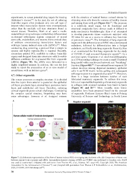
researchers printed PCL scaffolds to culture brain-like of COVID-19 and screened therapeutic agents . In a
[79]
organs and designed them into structures with favorable previous report, Grigoryan et al. used stereolithography
diffusion conditions for engineered flat brain organoids as a 3D bioprinting technique to create a small 3D printed
(efBOs) (Figure 7B). The efBOs were fabricated in a lung model with a multivessel network and “breathing”
highly simplified manner. In addition, this was the first function (Figure 8B) . Conventional breast organoid 3D
[80]
study to report the preparation of an in vitro model of culture involves mixing dispersed mammary epithelial
neural tissue with an intrinsic gyrus . cells in an ECM matrix before gelling and subsequent
[7]
self-organization into organoid structures [81,82] . However,
4.7. Other organoids there is a large variation between batches of such
The cornea possesses a complex structure. It is divided fabricated mammary organoids. To address this issue,
into five layers from anterior to posterior: the epithelial John et al. reported the bioprinting of mammary organoids
cell layer, preelastic layer, stromal layer, posterior elastic in collagen with minimal variation from batch to batch
layer, and endothelial cell layer. Therefore, culturing (Figure 8C and D) . More recently, more bionic
[83]
corneal organoids poses certain challenges. Considering assemblies have been proposed based on the concept
the complex spatial structure, bioprinting may have of organoids. Professor Kunyoo Shin’s team at Pohang
some advantages. Isaacson et al. designed corneas University of Science and Technology in South Korea
A B
Figure 7. (A) Schematic representation of the flow of human induced PSCs developing into forebrain cells. (B) Comparison of tissue core
between regular brain organoids and efBOs. Immunohistochemical staining of NES and TUBB3 was performed to visualize cells. DAPI
was used as counterstain. (Adapted with permission from Trevino AE, et al., Science, 367: eaay1645, Copyright 2020, The American
Association for the Advancement of Science ) and (Adapted with permission from Theresa S P Rothenbücher et al., Biofabrication,
[72]
2021,13 011001, IOP Publishing Ltd ).
[76]
30 International Journal of Bioprinting (2021)–Volume 7, Issue 3