
Page 35 - IJB-7-3
P. 35
Ren, et al
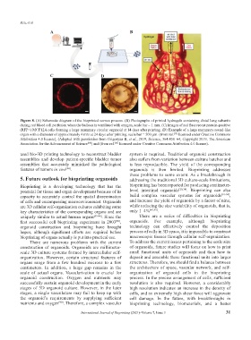
A B
C D
Figure 8. (A) Schematic diagram of the bioprinted cornea process. (B) Photographs of printed hydrogels containing distal lung subunits
during red blood cell perfusion when the balloon is ventilated with oxygen, scale bar = 1 mm. (C) Images of red fluorescent protein-positive
(RFP+) MCF12A cells forming a large mammary circular organoid at 14 days after printing. (D) Example of a large mammary round-like
organ with a diameter of approximately 4 mm at 24 days after printing, scale bar = 500 µm. (from ref. licensed under Creative Commons
[77]
Attribution 4.0 license), (Adapted with permission from Grigoryan B, et al., 2019, Science, 364:458–64, Copyright 2019, The American
Association for the Advancement of Science ) and (from ref. licensed under Creative Commons Attribution 4.0 license).
[83]
[80]
used bio-3D printing technology to reconstruct bladder system is required. Traditional organoid construction
assemblies and develop patient-specific bladder tumor also suffers from variation between culture batches and
assemblies that accurately mimicked the pathological is less reproducible. The yield of the corresponding
features of tumors in vivo . organoids is then limited. Bioprinting addresses
[84]
these problems to some extent. As a breakthrough in
5. Future outlook for bioprinting organoids addressing the traditional 3D culture-scale limitations,
Bioprinting is a developing technology that has the bioprinting has been reported for producing centimeter-
potential for tissue and organ development because of its level intestinal organoids [20,34] . Bioprinting can also
capacity to accurately control the spatial dissemination build complex vascular systems for organoids [91-94] ,
of cells and encompassing microenvironment. Organoids and increase the yield of organoids by a factor of nine,
are 3D cellular self-organization cultures exhibiting some while reducing the size variability of organoids, that is,
key characteristics of the corresponding organs and are only 1–4% [45,95] .
uniquely similar to actual human organs [85-88] . Since the There are a series of difficulties in bioprinting
first successful cell bioprinting experiment in 2003 , organoids. For example, although bioprinting
[89]
organoid construction and bioprinting have brought technology can effectively control the deposition
hopes, although significant efforts are required before process of cells in 3D space, it is impossible to construct
bioprinting of organs actually is put into practical use. macroscopic tissues through cellular self-organization.
There are numerous problems with the current To address the current issues pertaining to the scale size
construction of organoids. Organoids are millimeter- of organoids, future studies will focus on how to print
scale 3D culture systems formed by intercellular self- small functional units of organoids and then how to
organization. However, certain structural features of deposit and assemble these functional units into larger
organs range from a few hundred microns to a few structures. Therefore, we should find a balance between
centimeters. In addition, a large gap remains in the the architecture of space, vascular network, and self-
scale of actual organs. Vascularization is crucial for organization of organoid cells in the bioprinting
organoid construction. Oxygen and nutrients may process. In the precise arrangement of cells, sufficient
successfully sustain organoid development in the early resolution is also required. However, a considerably
stages of 3D organoid culture. However, in the later high resolution indicates an increase in the density of
stages, a single vasculature may fail to keep up with cells, and an extremely high shear force will aggravate
the organoid’s requirements by supplying sufficient cell damage. In the future, with breakthroughs in
nutrients and oxygen [90] . Therefore, a complex vascular bioprinting technology, biomaterials, and a better
International Journal of Bioprinting (2021)–Volume 7, Issue 3 31