
Page 31 - IJB-7-3
P. 31
Ren, et al
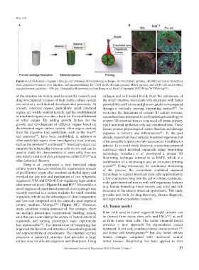
A B
Figure 4. (A) Schematic diagram of the pre-set extrusion 3D bioprinting technique for liver lobule printing. (B) MIX and preset structures
were compared to assess liver function, and immunostaining for CD31 (red), albumin (green), MRP2 (green), and DAPI cell nuclei (blue)
was performed; scale bar = 200 µm. (Adapted with permission from Kang et al, Small, Copyright 2020 Wiley-VCH Verlag ).
[51]
of the intestine are widely used in scientific research and collagen and cell-loaded bioink from the submucosa of
drug development because of their stable culture system the small intestine, microscale villi structures with better
and relatively well-defined developmental processes. At permeability coefficients and glucose uptake were prepared
present, intestinal organs, particularly small intestinal through a vertically moving bioprinting method . To
[60]
organs, are widely studied models, and the establishment overcome the limitations of current 3D culture systems,
of intestinal organs provides a basis for the establishment researchers have attempted to use bioprinting technology to
of other organs. By adding growth factors for the prepare 3D intestinal tissues composed of human primary
growth and development of different organs based on small intestinal epithelial cells and myofibroblasts. These
the intestinal organ culture system, other organs derived tissues possess physiological barrier function and damage
from the digestive tract epithelium, such as the liver response to toxicity and inflammation . In the past
[47]
[61]
and pancreas , have been established, in addition to decade, researchers have cultured intestinal organoids that
[53]
other epithelial organs from non-digestive tract sources, often assemble hepatocytes into micrometer to millimeter
such as the prostate and breast . Intestinal organs can spheres. In a recent study, however, researchers prepared
[54]
[55]
simulate the relationship between cells in vivo and can be centimeter-sized intestinal organoids using bioprinting
used to study the characteristics of stem cells; they are technology. Jonathan et al. developed a unique 3D
also widely used in studying ulcerative colitis (UC) [56] and bioprinting technique referred to as BATE, which is a
other intestinal diseases. combination of a microscope and an extrusion printing
Deng et al. constructed a new intestinal organ system . Using microscopy for continuous monitoring
[20]
culture system that can simulate the regeneration process of the process, the researchers combined organoid
of proliferative crypts after intestinal epithelial injury and technology to deposit intestinal stem cells approximately
revealed the key role and mechanism of two epigenetic a few centimeters long into the gel to obtain centimeter-
regulators (VPA and EPZ6438) in regulating regeneration scale gastrointestinal tissues with self-organizing features
after intestinal injury (Figure 5A and B) . Meanwhile, a (e.g. lumen, branching blood vessels, and crypt and villi
[57]
novel engineered plant-based nanocellulose hydrogel was structures of the tubular intestinal epithelium). This study
recently reported as a culture medium for small intestine provides new tools for drug discovery, disease diagnosis,
organoids, which has the advantages of clear composition and regenerative medicine research.
and low cost compared with the currently used organoid
culture medium, Matrigel (Figure 5C). However, 4.5. Tumor model
[58]
many questions remain unanswered. For example, there
are multiple phenotypes (symmetrical, budding, mixed, Stem cells used in tumor organoid model cultures can
[62]
etc.) that can occur during the culture of human intestinal be derived from tissue stem cells and PSCs , as well
organoids, and varying phenotypes make experiments as from tumor stem cells. The tumor organoid model
less reproducible . The introduction of bioprinting has provides a new approach for personalized cancer
[59]
improved the function and structure of intestinal organoids treatment. It not only simulates tumor characteristics [63]
and reproducibility of experiments. The intestinal surface and tumor cell heterogeneity but also better reflects
[62]
possesses a microvilli structure that provides a large human changes compared with traditional animal
surface area for efficient digestion and absorption. Using tumor models. Bioprinting has been applied to alter
International Journal of Bioprinting (2021)–Volume 7, Issue 3 27