
Page 28 - IJB-7-3
P. 28
3D Bioprinted Organoids
4.1. Heart culture in response to the classical Wnt signaling
The heart is one of the most important organs in the pathway, thereby effectively differentiating them into
human body, providing power to support the flow highly purified cardiomyocytes and establishing a 3D
of blood, supplying various nutrients and oxygen to heart-like structure of a certain size with different cell
[38]
other organs and tissues, and eliminating the waste layer patterns and a foreground endoderm structure .
products of metabolism so that the body can function However, the organoid thus constructed successfully
properly. A mature heart contains 9 billion cells, replicates some aspects of heart tissue, including stromal
including fibroblasts, cardiomyocytes, smooth muscle cells, endothelial cell network, and epicardial layer, and
cells, connective tissue cells, and immune cells [32,33] . even resembles to early heart developmental morphology,
Furthermore, unlike other parts of the body, the heart but the macroscopic structure of the organoid moderately
tissue cannot heal itself from damage. The current differs from that of the real organ. The incorporation of
challenge faced in bioprinting cardiac organs is that bioprinting has shown satisfactory results. In April 2019,
the biomaterials used in bioprinting cardiac organs are scientists in Israel successfully 3D printed an “artificial
primarily the soft materials that possess low mechanical heart,” which is the first successfully designed and
strength and weak support, making it difficult to print printed heart comprising cells, blood vessels, ventricles,
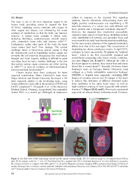
and shape layer by layer. Another challenge is the fact and atria (Figure 2A, B and C). Although the cells in
that current cardiac organ constructs are either lacking this heart appear to contract, they cannot beat and pump
[36]
in cells [34,35] or have no evidence of electromechanical blood like a normal heart . Recently, Professor Adam
function and lack functionality . Feinberg’s team at Carnegie Mellon University used
[36]
Certain studies have been conducted for heart collagen as a freely embeddable suspension hydrogel
organoid construction. Ishino Fumitoshi’s team from (FRESH) to bioprint heart organoids, combining MRI
Tokyo Medical and Dental University developed a 3D images of coronary arteries and 3D images of the heart
heart organoid, similar to the developing heart, using to achieve fine structures at different structural scales
mouse embryonic stem cells in the presence of FGF4 and from capillaries to the entire heart organ, as well as
LN/ET complexes . Zweigerdt et al. of the Hannover high-resolution printing of heart organoids with systolic
[37]
Medical School, Germany, encapsulated free-suspended function. (Figure 2D, E, and F). Previously constructed
[34]
human PSCs in a matrix gel (Matrigel) in suspension organoids are almost always millimeter-sized. However,
A B C
D E F
Figure 2. (A) Side view of the bioprinting concept and the unique cellular bioink. (B) 3D confocal image of a bioprinted heart (CM in
pink, EC in orange), scale bar =1 mm. (C) Bioprinted heart in a support bath. (D) A schematic diagram of fast cross-linking by squeezing
the collagen solution in a support bath with a pH of 7.4. (E and F) Magnetic resonance imaging (MRI) post-processed images of the heart
model, showing it has a coronary vascular network. (Adapted with permission from Lee et al, Science, 2019, 482–487 (2019) ) and (from
[34]
ref. licensed under Creative Commons Attribution 4.0 license).
[36]
24 International Journal of Bioprinting (2021)–Volume 7, Issue 3