
Page 32 - IJB-7-3
P. 32
3D Bioprinted Organoids
A
B
C
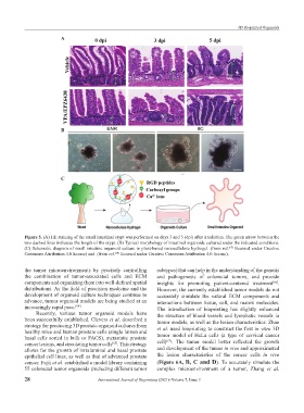
Figure 5. (A) HE staining of the small intestinal crypt was performed on days 3 and 5 (dpi) after irradiation. The green arrow between the
two dashed lines indicates the length of the crypt. (B) Typical morphology of intestinal organoids cultured under the indicated conditions.
(C) Schematic diagram of small intestine organoid culture in plant-based nanocellulose hydrogel. (from ref. licensed under Creative
[57]
Commons Attribution 4.0 license) and (from ref. licensed under Creative Commons Attribution 4.0 license).
[58]
the tumor microenvironments by precisely controlling subtypes)that can help in the understanding of the genesis
the combination of tumor-associated cells and ECM and pathogenesis of colorectal tumors, and provide
components and organizing them into well-defined spatial insights for promoting patient-centered treatment .
[66]
distributions. As the field of precision medicine and the However, the currently established tumor models do not
development of organoid culture techniques continue to accurately simulate the natural ECM components and
advance, tumor organoid models are being studied at an interactions between tissue, cell, and matrix molecules.
increasingly rapid pace. [64] The introduction of bioprinting has slightly enhanced
Recently, various tumor organoid models have the structure of blood vessels and lymphatic vessels in
been successfully established. Clevers et al. described a tumor models, as well as the lesion characteristics. Zhao
strategy for producing 3D prostate organoid cultures from et al. used bioprinting to construct the first in vitro 3D
healthy mice and human prostate cells (single lumen and
basal cells sorted in bulk or FACS), metastatic prostate tumor model of HeLa cells (a type of cervical cancer
[67]
cancer lesions, and circulating tumor cells . This strategy cell) . The tumor model better reflected the growth
[65]
allows for the growth of intraluminal and basal prostate and development of the tumor in vivo and approximated
epithelial cell lines, as well as that of advanced prostate the lesion characteristics of the cancer cells in vivo
cancer. Fujii et al. established a model library containing (Figure 6A, B, C and D). To accurately simulate the
55 colorectal tumor organoids (including different tumor complex microenvironment of a tumor, Zhang et al.
28 International Journal of Bioprinting (2021)–Volume 7, Issue 3