
Page 50 - IJB-7-3
P. 50
3D Bioprinting Photo-crosslinkable Hydrogels for Bone and Cartilage Repair
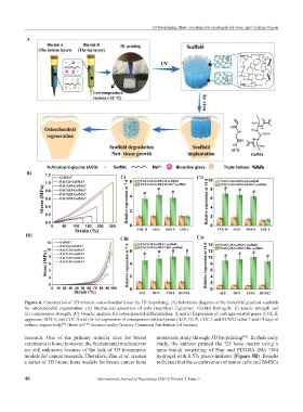
A A
Bi
Ci Cii
Bii
Ciii Civ
Figure 4. Construction of 3D mimetic osteochondral tissue by 3D bioprinting. (A) Schematic diagram of the biohybrid gradient scaffolds
for osteochondral regeneration. (B) Mechanical properties of poly (nacryloyl 2-glycine) -GelMA hydrogels: (i) tensile strength and
(ii) compressive strength. (C) Genetic analysis for osteochondral differentiation: (i and ii) Expression of cartilage-related genes (COL II,
aggrecan, SOX-9, and COL I) and (iii-iv) expression of osteogenesis-related genes (ALP, OCN, COL I, and RUNX2) after 7 and 14 days of
culture, respectively (from ref. licensed under Creative Commons Attribution 4.0 license).
[79]
[79]
research. One of the primary transfer sites for breast metastasis study through 3D bioprinting . In their early
[85]
carcinoma is bone; however, the fundamental mechanisms study, the authors printed the 3D bone matrix using a
are still unknown because of the lack of 3D biomimetic nano-bioink consisting of Hap and PEGDA (Mn 700)
models for cancer research. Therefore, Zhu et al. created hydrogel with 0.5% photo-initiator (Figure 5B). Results
a series of 3D bionic bone models for breast cancer bone indicated that the cocultivation of tumor cells and BMSCs
46 International Journal of Bioprinting (2021)–Volume 7, Issue 3