
Page 52 - IJB-7-3
P. 52
3D Bioprinting Photo-crosslinkable Hydrogels for Bone and Cartilage Repair
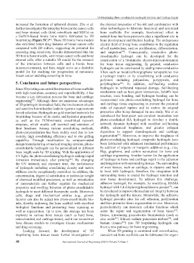
increased the formation of spheroid clusters. Zhu et al. the physical properties of bio-ink and combination with
further investigated the interplay between the cancer cells other technologies to fabricate functional and optimized
and bone stromal cells (fetal osteoblasts and MSCs) on bone scaffolds. For example, bioelectrical effect in
a GelMA-based bionic bone matrix fabricated by 3D natural bone has been proven to play a significant role in
bioprinting (Figure 5C) . These 3D bionic bone models bone development and fracture healing. The endogenous
[86]
also exhibited better drug fastness of breast cancer cells electric field of living bone contributes to the regulation
compared with 2D culture, suggesting its potential for of cell metabolism, such as proliferation, differentiation,
assessing drug sensitivity. Results demonstrated that the and migration . Consequently, conductive photo-
[89]
3D bionic bone models, with breast cancer cells and bone crosslinkable hydrogel can be developed for the
stromal cells, offer a suitable 3D model for the research construction of a biomimetic electro-microenvironment
of the interaction between cells and a bionic bone for bone tissue engineering. In general, conductive
microenvironment, and thus, might serve as a desirable hydrogels can be constructed by adding nano-conductive
model for the studying the progression of metastatic fillers such as metal/carbon/graphene nanoparticles into
breast cancer and drug screening in bone. a hydrogel matrix or by crosslinking with conductive
polymers including polyaniline, polypyrrole, and
5. Conclusion and future perspectives polythiophene . In addition, self-healing enables
[90]
Since 3D printing can control the structure of tissue scaffolds hydrogels to withstand repeated damage. Self-healing
with high resolution, accuracy and reproducibility, it has mechanism such as host-guest interactions, Schiff’s base
become a key fabrication process for bone and cartilage reaction, ionic bonding, and hydrogen bonding can be
engineering . Although there are numerous advantages incorporated into photo-crosslinking hydrogel for bone
[87]
of 3D printing in biomedical field, the involvement of cells and cartilage tissue engineering to prevent the potential
and sensitive biomolecules leads to rigorous requirements risks of repeated rupture and to restore its original
for ink selection. Hydrogels are gold standard materials for properties after being damaged. In one study, Wei et al.
bioprinting because of its elastic and hydrated properties introduced the host-guest non-covalent interaction into
as well as the ECM-mimetic crosslinked network photo-crosslinked HA hydrogel to develop a double
structure, which enable cell survival and retention of network dynamic hydrogel with excellent self-healing
their functions. Among various crosslinking methods, ability, enhanced toughness, and greater collagen
photo-polymerization has been widely used due to low deposition to support chondrogenesis and cartilage
[91]
toxicity, high crosslinking efficiency and in situ gelling regeneration . Moreover, to improve the toughness of
capability. Through modification by computer-aided photo-crosslinking hydrogel, composite hydrogels have
design/manufacturing or medical imaging systems, photo- been fabricated with enhanced mechanical performance
crosslinkable hydrogels can be personalized at different by addition of organic or inorganic additives (e.g., clay,
bone length scale by 3D printing. With the application of Hap, grapheme, and carbon nanotubes) for bone and
UV light, the photo-crosslinking hydrogels undergo rapid cartilage engineering. Another barrier for the application
formation immediately after printing . By changing of hydrogel to bone and cartilage repair is the adhesion
[88]
the UV intensity and exposure time, the performance and integration with surrounding tissues. The surrounding
of hydrogels including crosslinking density and matrix of most tissues, such as cartilage, is slippery and hard
stiffness can be exceptionally controlled. In addition, the to bond with hydrogels; therefore, the integration with
concentration, degree of substitution or molecular weight surrounding tissue is crucial for hydrogel retention and
of chemical modified precursors, as well as introduction new tissue development. To address this challenge,
of nanomaterials can further regulate the mechanical adhesive hydrogel, for example, by modifying HAMA
properties and swelling behavior of photo-crosslinkable hydrogel with 3,4-dihydroxyphenylalanine groups , can
[92]
hydrogels to meet different therapeutic needs. Moreover, be developed to improve the mechanical integrity between
cells, drugs and biochemical entities (e.g., growth the hydrogels and the tissues. Meanwhile, the adhesive
factors) can also be added into photo-crosslinkable bio- hydrogel provides sites for cell adhesion, proliferation
inks, thereby endowing the bone scaffold with excellent and thus promotes tissue regeneration in vivo. Moreover,
biological functions and promoting bone repairing as piezoelectricity can modulate cellular functions which
well as regeneration. Up to now, this strategy has been assist the repair and regeneration of bone tissue .
[93]
explored in various bone tissues (such as hard bone, Hence, introducing piezoelectric biomaterials (such as
osteochondral, and cartilage tissue), and it can reconstruct zinc oxide , lithium sodium potassium niobate , and
[93]
[94]
bone disease models to investigate disease mechanisms barium titanate ) into 3D bioprinting hydrogels can
[95]
and drug screening. blaze a new pathway for bone engineering.
Looking forward, the development of 3D When 3D printing is combined with microfluidics,
bioprinting bone tissues needs further investigation of vascularized bone scaffolds can be fabricated for organ-
48 International Journal of Bioprinting (2021)–Volume 7, Issue 3