
Page 214 - IJB-8-1
P. 214
Nano-Hydroxyapatite Bone Scaffolds with Different Porous Structures Processed by Digital Light Processing 3D Printing
optimized parameters. The compressive properties of zirconia beads were added into the slurry. After
and in vitro biological evaluation in cell proliferation vigorously mixing in a Turbula shaker-mixer (Turbula
and attachment morphologies of three structures were T2F, Basel, Switzerland) for above 6 h, the nano-HA
compared and studied. Our research is expected to offer slurry was obtained.
an insight into guide the bioceramic scaffold fabrication
and the selection for BTE applications. 2.3. Design and fabrication of scaffolds
2. Materials and methods The TPMS-P surface belongs to a member of the TPMS
family, which can be defined by the specific mathematical
2.1. Materials equation [26,27] , referring to Equation (2.1) where is defined
as unit size and represents the expansion of surface. In
The HA powders (diameter: 20 nm; length: 270 nm) this work, the P structure of scaffolds was designed by
were supplied by Nanjing Emperor Nano Materials Co., MATLAB.
Ltd (Jiangsu, China); 1, 6-hexanediol diacrylate (HDDA,
Shanghai Yinchang Materials Co., China) was selected as Φ ( xy z,, ) = cos( x) + cos( y) + cos( z) = (2.1)
c
p
the monomer of the slurry. Its two reactive functionalities
ensure a sufficient cross-linking in curing . The dispersant- BCC and CPS structures with symmetric features
[23]
BYK for powder surface modification was provided by were designed by CAD software. All scaffolds were
BYK Additive and Instrument, Germany. TPO (Diphenyl designed with the same porosity value, that is, ~65%.
(2, 4, 6-trimethylbenzoyl)) phosphine oxide from Shanghai The scaffolds were printed by a top-down DLP 3D
Macklin Biochemical Co. was used as photoinitiator. 3-[4, printer (405 nm light source) with a 30 um layer thickness
5-dimethylthiazol-2-yl]-2, 5-diphenyltetrazolium bromide (~10 mJ/cm energy dose, 1.5 s exposure time). First, the
2
(MTT) was provided by Invitrogen (Thermal Fisher as-prepared nano-HA slurry was poured into the tank
Scientific, USA). Dulbecco’s Modified Eagle’s Medium until the volume of slurry can meet the need of printing
(DMEM), fetal bovine serum, and penicillin-streptomycin models. The 3D models were sliced into 2D images by
were provided by Gibco (Thermal Fisher Scientific, USA). a slicing software. Basing on the slicing data, the slurry
Paraformaldehyde (PFA) was supplied by Alfa Aesar. was selectively cured by ultraviolet light layer by layer
2.2. Slurry preparation till the printing was fully accomplished. After finishing
the printing process, the green parts were immersed in a
Before slurry preparation, surface modification of the mixture consisting of monomers and ethanol, and washed
nano-HA powders is necessary for homogeneous and by an ultrasonic cleaning to remove the residual slurry.
stable dispersion [24,25] . The modification process is To determine the debinding and sintering strategy,
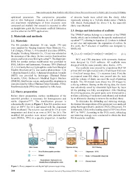
schematically shown in Figure 1. Raw HA powders were the thermal decomposition of the green part was analyzed
dried at 100°C for 12 h and mixed with the dispersant using thermogravimetric analysis (TGA) (Discovery, TA
BYK in ethanol. The solution was stirred for 2 h at room Instruments, USA) with a heating speed of 10°C/min from
temperature and dried in an oven at 50°C for 24 h. The 40°C to 700°C. According to the TGA result, debinding
modified HA powders were mixed with photoinitiator and sintering strategy were conducted to obtain the final
resin (HDDA, TPO) in a specific proportion. A number HA bioceramic scaffolds.
Figure 1. Slurry preparation and digital light processing printing.
200 International Journal of Bioprinting (2022)–Volume 8, Issue 1