
Page 217 - IJB-8-1
P. 217
Liang, et al.
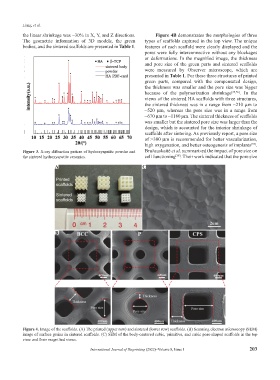
the linear shrinkage was ~30% in X, Y, and Z directions. Figure 4B demonstrates the morphologies of three
The geometric information of 3D models, the green types of scaffolds captured in the top view. The unique
bodies, and the sintered scaffolds are presented in Table 1. features of each scaffold were clearly displayed and the
pores were fully interconnective without any blockages
or deformations. In the magnified image, the thickness
and pore size of the green parts and sintered scaffolds
were measured by Observer microscope, which are
presented in Table 1. For these three structures of printed
green parts, compared with the compensated design,
the thickness was smaller and the pore size was bigger
because of the polymerization shrinkage [35,36] . In the
views of the sintered HA scaffolds with three structures,
the sintered thickness was in a range from ~210 µm to
~520 µm, whereas the pore size was in a range from
~670 µm to ~1180 µm. The sintered thickness of scaffolds
was smaller but the sintered pore size was larger than the
design, which is accounted for the interior shrinkage of
scaffolds after sintering. As previously report, a pore size
of >300 µm is recommended for better vascularization,
high oxygenation, and better osteogenesis of implants .
[37]
Figure 3. X-ray diffraction pattern of hydroxyapatite powder and Bružauskaitė et al. summarized the impact of pore size on
[38]
the sintered hydroxyapatite ceramics. cell functioning . Their work indicated that the pore size
A C
B
Figure 4. Image of the scaffolds. (A) The printed (upper row) and sintered (lower row) scaffolds. (B) Scanning electron microscopy (SEM)
image of surface grains in sintered scaffolds. (C) SEM of the body-centered cubic, primitive, and cubic pore-shaped scaffolds in the top
view and their magnified views.
International Journal of Bioprinting (2022)–Volume 8, Issue 1 203