
Page 219 - IJB-8-1
P. 219
Liang, et al.
A B
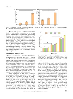
Figure 5. Mechanical properties of body-centered cubic, primitive, and cubic pore-shaped scaffolds. (A) Compressive strength.
(B) Compressive modulus. * represents P < 0.05.
Modulus of the scaffolds is another key element that
should be analyzed. The mismatching of the modulus
between native bone and implants could result in stress
shielding, thus leading to the original bone loss .
[42]
Figure 5B shows the compressive modulus of these three
types of scaffolds. The CPS scaffold still performed the
highest modulus, ~400 MPa, and significantly higher than
the P and BCC scaffolds. The compressive strength and
modulus of cancellous bone range from 1.6 MPa to 4.6
MPa and from 22.9 MPa to 431 MPa, respectively [43-45] .
All scaffolds with different structures designed in this
study exhibited mechanical properties that are comparable
with the native cancellous bone, indicating their potential
in bone applications.
3.4. Biological testing in vitro
MTT assay was conducted to evaluate the cell proliferation Figure 6. 3-[4, 5-Dimethylthiazol-2-yl]-2, 5-diphenyltetrazolium
of rBMSCs cultured onto the P, BCC, and CPS scaffolds bromide assay for proliferation of rat bone mesenchymal stem
for 1, 4, and 7 days. The increasing absorbance of three cells culturing for 1, 4, and 7 days on three scaffolds. * represents
scaffolds can be seen with the days of cultivation in P < 0.05.
Figure 6, indicating that HA scaffolds formed through
the mentioned fabrication process were non-cytotoxic majority of rBMSCs obviously exhibited the spreading
and biocompatible. After 7 days cultivation, the cell behavior on day 4. This suggests that cell metabolism
viability was generally more than ~75% compared with on day 4 was more active than that on day 1, which
the control group. Meanwhile, the absorbance of both was in good agreement with the MTT result on day
BCC and CPS scaffolds was significantly higher than 4, as shown in Figure 6. On day 7, the cell spread of
that of P scaffolds, which revealed that BCC and CPS rBMSCs became larger and they formed a thin membrane
scaffolds were beneficial for cell metabolisms. to cover the scaffolds. The similar processes of the cell
Figure 7B-D shows the SEM images of rBMSCs’ membrane covering the scaffolds have been reported [56,57] .
morphologies after being cultured for 1, 4, and 7 days This indicates the active attachment of rBMSCs to the
on the three types of scaffolds. On day 1, the rBMSCs scaffolds. Meanwhile, SEM images of day 7 showed that
generally adhered well and maintained the spindle-like the rBMSCs deposited the extracellular matrix (ECM) on
morphology, confirming the non-cytotoxicity of the all scaffolds. Particularly, the amount of ECM formed on
scaffolds [46-51] . In magnified images (Figure 7A), the the surface of CPS scaffolds was obviously higher than
filopodia adhering on the surface of scaffolds could be that of others. ECM plays a critical role in providing
seen clearly, indicating a good cell attachment. Cell support for cell growth and migration . It may exhibit
[38]
spreading is a sign of adherence to a substrate, which the metabolism of rBMSCs which are more active on
directly or indirectly regulates the cell metabolism [52-55] . CPS scaffolds, which can explain the highest absorbance
On three types of scaffolds, compared with day 1, the at day 7 in Figure 6.
International Journal of Bioprinting (2022)–Volume 8, Issue 1 205