
Page 125 - IJB-8-3
P. 125
Zheng, et al.
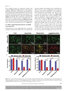
PGA scaffolds showed no cytotoxicity without NIR sterilized scaffolds and irradiated with or without 808 nm
irradiation (Figure 5A and B). However, under NIR laser NIR for 10 min. As show in Figure 6A, under NIR laser
irradiation, MoS /PGA, PMoS /PGA, and Ag@PMoS / irradiation, MoS /PGA, PMoS /PGA, and Ag@PMoS /
2
2
2
2
2
2
PGA scaffolds significantly ablated MG63 cells. The area PGA scaffolds also ablated BMSC cells. The area ratio
occupied by living cells decreased on average by 8.4%, of living cells decreased by about 2% (Figure 6B).
which was significant superior to the PGA scaffolds alone However, the ablation effect of these scaffolds on MSC
(Figure 5A and B). More importantly, compared with cells was significantly reduced compared with those on
MoS /PGA, the ablation effect of Ag@PMoS /PGA on MG63 cells. As shown in Figure 5C and 6C, two kinds
2
2
tumor cells was significantly enhanced (Figure 5A and B). of cells were cocultured with the composite scaffold,
Cell Counting Kit-8 also showed that the survival rate of respectively, the activity of MG63 cells was about 0.5,
MG63 cells decreased significantly by 39% (Figure 5C). while that of BMSC cells was still about 1.4 after NIR
3.5. Effect of photothermal function of scaffold irradiation. This is because tumor cells are far less
on normal cells tolerant to heat than normal cells and are easily killed
by high temperature. When the tumor tissue was heated
At the same time, we also evaluated the effect of composite at about 43℃, the permeability of tumor cell membrane
scaffolds on normal cells. BMSC cells were cultured with was increased. When the temperature is above 50℃, the
A
B C
Figure 5. In vitro photothermal antitumor role of the composite scaffolds fabricated by SLS. Live/dead staining (A), statistical diagram of
living cell proportion area (B), and Cell Counting Kit-8 assay (C) of MG63 cells after incubation with PGA, MoS /PGA, PMoS /PGA, and
2
2
Ag@PMoS /PGA scaffolds with or without 808 nm NIR irradiation (1 W/cm , 10 min).
2
2
International Journal of Bioprinting (2022)–Volume 8, Issue 3 117
Please cite this article as: Zheng L, Zhong Y, He T, et al., 2022, A Codispersed Nanosystem of Silver-anchored MoS Enhances Antibacterial
2
and Antitumor Properties of Selective Laser Sintered Scaffolds, Int J Bioprint, 8(3):0025. http://doi.org/10.18063/ijb.v8i3.0025