
Page 124 - IJB-8-3
P. 124
3D Scaffold for Combined Antibacterial and Antitumor Therapy
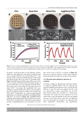
Figure 4. The microstructure and photothermal performance of scaffolds. (A-D) Photographs and (A1-D1) SEM images of the cross-section
of the composite scaffolds. (E) Real-time temperature of PGA, MoS /PGA, PMoS /PGA, and Ag@PMoS /PGA scaffolds during NIR
2
2
2
irradiation. (F) Real-time temperature of Ag@PMoS /PGA scaffolds under “on-off” cycles during the NIR irradiation.
2
In general, scaffolds suitable for cell adhesion, growth, laser on/off cycles (1 W/cm ). As shown in Figure 4F,
2
migration, and appropriate mechanical strength should there was no obvious reduction, which further indicates
have an aperture between 100 μm and 1000 μm [40-43] . The that the photothermal agent has excellent photostability.
aperture of the composite scaffolds prepared in this study
was about 500 um, which was within a reasonable range. 3.4. Photothermal antitumor function of
Figure 4A1-D1 shows the SEM images of the cross section scaffolds
of the composite scaffolds fractured by liquid nitrogen. It
is obvious that there are some aggregates from the MoS / To further confirm the photothermal antitumor efficacy
2
PGA matrix due to the strong van der Waals forces of of composite scaffolds, MG63 cells were cultured with
MoS NSs . Conversely, the Ag@PMoS NSs were sterilized scaffolds and irradiated with or without 808 nm
[44]
2
2
uniformly distributed in the PGA matrix, which benefited NIR for 10 min. The cells were stained with PI in red
from the synergistic dispersion effect of the Ag NPs and and calcein-AM in green, respectively, and the live/
MoS NSs . Furthermore, we verify the photothermal dead staining status was evaluated by a fluorescence
[45]
2
performance of composite scaffolds by irradiating 808 nm microscope. The dead cells were stained in red as PI
NIR laser. As shown in Figure 4E, Ag@PMoS /PGA cannot enter living cells and the living cells were stained
2
scaffolds exhibited better photothermal performance than in green (calcein-AM enters dead cells and dispersed
PMoS /PGA and MoS /PGA. Furthermore, photothermal very fast and cannot be detected). As expected, both
2
2
stability of Ag@PMoS /PGA was examined by over four PGA, MoS /PGA, PMoS /PGA, and Ag@PMoS /
2
2
2
2
116 International Journal of Bioprinting (2022)–Volume 8, Issue 3
Please cite this article as: Zheng L, Zhong Y, He T, et al., 2022, A Codispersed Nanosystem of Silver-anchored MoS Enhances Antibacterial
2
and Antitumor Properties of Selective Laser Sintered Scaffolds, Int J Bioprint, 8(3):0025. http://doi.org/10.18063/ijb.v8i3.0025