
Page 128 - IJB-8-3
P. 128
3D Scaffold for Combined Antibacterial and Antitumor Therapy
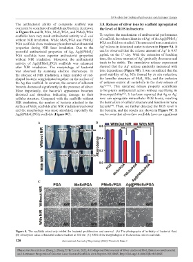
The antibacterial ability of composite scaffold was 3.8. Release of silver ions by scaffold upregulated
evaluated by coculture of scaffolds and bacteria. As shown the level of ROS in bacteria
in Figure 8A and B, PGA, MoS /PGA, and PMoS /PGA
2
2
scaffolds have very weak antibacterial activity to E. coli To explore the mechanism of antibacterial performance
+
without NIR irradiation. While MoS /PGA and PMoS / of scaffold, the release kinetics of Ag of the Ag@PMoS /
2
2
2
PGA scaffolds show moderate photothermal antibacterial PGA scaffold was studied. The amount of non-cumulative
+
properties during NIR laser irradiation. Due to the Ag release in deionized water is shown in Figure 9A. It
+
powerful antibacterial properties of Ag, Ag@PMoS / can be observed that the release amount of Ag is 0.87
2
st
PGA scaffolds have superior antibacterial properties μg/mL on the 1 day. With the extension of leaching
+
without NIR irradiation. Moreover, the antibacterial time, the release amount of Ag gradually decreases and
activity of Ag@PMoS /PGA scaffolds was enhanced tends to be stable. The cumulative release experiment
2
+
after NIR irradiation. The morphology of bacterial showed that the Ag release gradually increased with
was observed by scanning electron microscope. In time dependence (Figure 9B). It was considered that the
the absence of NIR irradiation, a large number of rod- good stability of Ag NPs formed by in situ reduction,
shaped bacteria conglutinated together on the surface of the lamellar structure of MoS NSs, and the inclusion
2
the Ag-free scaffold. In contrast, the content of adherent of polymer matrix all contribute to the slow release of
bacteria decreased significantly in the presence of silver. Ag +[58-61] . This sustained release property contributes
More importantly, the bacteria’s appearance becomes to long-term antibacterial action without sacrificing its
distorted and shrunken, indicating damage to their biocompatibility . It has been reported that Ag or Ag
[62]
+
cellular structure. Compared with the scaffolds without ions can upregulate intracellular ROS levels, resulting
NIR irradiation, the number of bacteria attached to the the destruction of cellular structure and function in many
surface of MoS scaffolds after NIR irradiation was lower bacteria . Then, we further detected the ROS level in
[63]
2
and the morphology was more atrophied, especially the the bacteria, and the results are shown in Figure 9C. It
Ag@PMoS /PGA scaffolds (Figure 8C). can be seen that silver-free scaffolds have no significant
2
A B
C
Figure 8. The scaffolds selectively inhibit the bacterial proliferation and survival. (A) The photographs of turbidity of bacterial fluid.
(B) Absorption value of bacterial culture medium at 600 nm. (C) SEM of the morphologies of Escherichia coli on scaffolds.
120 International Journal of Bioprinting (2022)–Volume 8, Issue 3
Please cite this article as: Zheng L, Zhong Y, He T, et al., 2022, A Codispersed Nanosystem of Silver-anchored MoS Enhances Antibacterial
2
and Antitumor Properties of Selective Laser Sintered Scaffolds, Int J Bioprint, 8(3):0025. http://doi.org/10.18063/ijb.v8i3.0025