
Page 126 - IJB-8-3
P. 126
3D Scaffold for Combined Antibacterial and Antitumor Therapy
A
B C
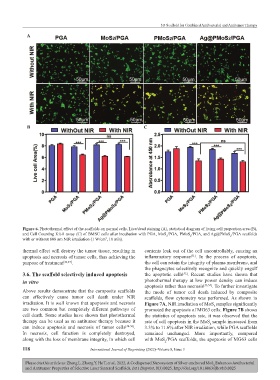
Figure 6. Photothermal effect of the scaffolds on normal cells. Live/dead staining (A), statistical diagram of living cell proportion area (B),
and Cell Counting Kit-8 assay (C) of BMSC cells after incubation with PGA, MoS /PGA, PMoS /PGA, and Ag@PMoS /PGA scaffolds
2
2
2
with or without 808 nm NIR irradiation (1 W/cm , 10 min).
2
thermal effect will destroy the tumor tissue, resulting in contents leak out of the cell uncontrollably, causing an
apoptosis and necrosis of tumor cells, thus achieving the inflammatory response . In the process of apoptosis,
[51]
purpose of treatment [46,47] . the cell can retain the integrity of plasma membrane, and
the phagocytes selectively recognize and quickly engulf
3.6. The scaffold selectively induced apoptosis the apoptotic cells . Recent studies have shown that
[52]
in vitro photothermal therapy at low power density can induce
apoptosis rather than necrosis [53,54] . To further investigate
Above results demonstrate that the composite scaffolds the mode of tumor cell death induced by composite
can effectively cause tumor cell death under NIR scaffolds, flow cytometry was performed. As shown in
irradiation. It is well known that apoptosis and necrosis Figure 7A, NIR irradiation of MoS samples significantly
2
are two common but completely different pathways of promoted the apoptosis of MG63 cells. Figure 7B shows
cell death. Some studies have shown that photothermal the statistics of apoptosis rate, it was observed that the
therapy can be used as an antitumor therapy because it rate of cell apoptosis in the MoS sample increased from
2
can induce apoptosis and necrosis of tumor cells [48-50] . 3.3% to 11.6% after NIR irradiation, while PGA scaffolds
In necrosis, cell function is completely destroyed, remained unchanged. More importantly, compared
along with the loss of membrane integrity, in which cell with MoS /PGA scaffolds, the apoptosis of MG63 cells
2
118 International Journal of Bioprinting (2022)–Volume 8, Issue 3
Please cite this article as: Zheng L, Zhong Y, He T, et al., 2022, A Codispersed Nanosystem of Silver-anchored MoS Enhances Antibacterial
2
and Antitumor Properties of Selective Laser Sintered Scaffolds, Int J Bioprint, 8(3):0025. http://doi.org/10.18063/ijb.v8i3.0025