
Page 91 - IJB-8-3
P. 91
Hu, et al.
The spine is a complex skeletal anatomic structure 2.2. 3D-printed titanium vertebral body design
involved in weight bearing, shock absorption, and motion. and manufacturing
Tumors involving the spine, whether primary or metastatic,
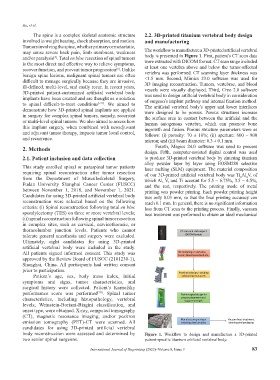
may cause severe back pain, limb numbness, weakness The workflow to manufacture a 3D-printed artificial vertebral
and/or paralysis . Total en bloc resection of spinal tumors body is presented in Figure 1. First, patient’s CT scan data
[5]
is the most direct and effective way to relieve symptoms, were extracted with DICOM format. CT scan range included
recover function, and prevent tumor progression . Unlike at least one vertebra above and below the tumor-affected
[6]
benign spine lesions, malignant spinal tumors are often vertebra was performed. CT scanning layer thickness was
<1.5 mm. Second, Mimics 23.0 software was used for
difficult to manage surgically because they are invasive, 3D imaging reconstruction. Tumors, vertebrae, and blood
ill-defined, multi-level, and easily recur. In recent years, vessels were visually displayed. Third, Creo 2.0 software
3D-printed patient-customized artificial vertebral body was used to design artificial vertebral body in consideration
implants have been created and are thought as a solution of surgeon’s implant pathway and internal fixation method.
to spinal difficult-to-treat conditions [7-9] . We aimed to The artificial vertebral body’s upper and lower interfaces
demonstrate how 3D-printed spinal implants are applied were designed to be porous. Porous structures increase
in surgery for complex spinal tumors, namely, recurrent the surface area in contact between the artificial and the
or multi-level spinal tumors. We also aimed to assess how human autogenous vertebra, which can promote bone
this implant surgery, when combined with neoadjuvant ingrowth and fusion. Porous structure parameters were as
and adjuvant tumor therapy, impacts tumor local control, follows: (i) porosity: 70 ± 10%; (ii) aperture: 600 – 800
and recurrence. micron; and (iii) beam diameter: 0.3 ± 0.1 mm.
2. Methods Fourth, Magics 24.0 software was used to process
design. Fifth, computer-assisted digital control was used
2.1. Patient inclusion and data collection to produce 3D-printed vertebral body by sintering titanium
alloy powder layer by layer using EOSM280 selective
This study enrolled spinal or paraspinal tumor patients laser melting (SLM) equipment. The material composition
requiring spinal reconstruction after tumor resection of our 3D-printed artificial vertebral body was Ti Al V, of
6
4
from the Department of Musculoskeletal Surgery, which Al, V, and Ti account for 5.5 – 6.75%, 3.5 – 4.5%,
Fudan University Shanghai Cancer Center (FUSCC) and the rest, respectively. The printing mode of metal
between November 1, 2018, and November 1, 2021. printing was powder printing. Each powder printing height
Candidates for using 3D-printed artificial vertebral body was only 0.03 mm, so that the final printing accuracy can
reconstruction were selected based on the following reach 0.1 mm. In general, there is no significant information
criteria: (i) Spinal reconstruction following total en bloc loss from CT scan to the printing process. Finally, vacuum
spondylectomy (TES) on three or more vertebral levels; heat treatment was performed to obtain an ideal mechanical
(ii) spinal reconstruction following spinal tumor resection
in complex sites, such as cervical, cervicothoracic, or
thoracolumbar junction levels. Patients who cannot
tolerate general anesthesia and surgery were excluded.
Ultimately, eight candidates for using 3D-printed
artificial vertebral body were included in the study.
All patients signed informed consent. This study was
approved by the Review Board of FUSCC (2101230-1),
Shanghai, China. All participants had written consent
prior to participation.
Patient’s age, sex, body mass index, initial
symptoms and signs, tumor characteristics, and
surgical history were collected. Patient’s Karnofsky
performance score was performed [10] . Spinal tumor
characteristics, including histopathology, vertebral
levels, Weinstein-Boriani-Biagini classification, and
onset type, were obtained. X-ray, computed tomography
(CT), magnetic resonance imaging, and/or positron
emission tomography (PET)-CT were scanned. All
candidates for using 3D-printed artificial vertebral
body reconstruction were assessed and determined by Figure 1. Workflow to design and manufacture a 3D-printed
two senior spinal surgeons. patient-specific titanium artificial vertebral body.
International Journal of Bioprinting (2022)–Volume 8, Issue 3 83