
Page 96 - IJB-8-3
P. 96
3D-Printed Artificial Vertebral Body
A B A B
C
C
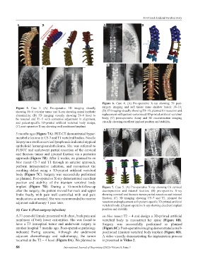
Figure 6. Case 4. (A) Pre-operative X-ray showing T9 post-
Figure 5. Case 3. (A) Pre-operative 3D imaging visually surgery imaging and soft tissue mass shadow beside T8~10;
showing T6~8 reticular tumor and X-ray showing spinal kyphotic (B) 3D imaging visually showing T8~10, planned for resection and
abnormality; (B) 3D imaging visually showing T6~8 level to replacement with patient-customized 3D-printed artificial vertebral
be resected and T1~5 with corrective adjustment in alignment, body; (C) post-operative X-ray and 3D reconstruction imaging
and patient-specific 3D-printed artificial vertebral body design; visually showing excellent implant position and stability.
(C) post-operative X-ray showing well positioned implant.
A B
5 months ago (Figure 7A). PET-CT demonstrated hyper-
metabolic lesions in C5-7 and T1 vertebral bodies. Needle
biopsy on a swollen cervical lymph node indicated atypical
epithelioid hemangioendothelioma. She was referred to
FUSCC and underwent partial resection of the cervical
and thoracic tumor and internal fixation via a posterior
approach (Figure 7B). After 2 weeks, we planned to en C D
bloc resect C5-7 and T1 through an anterior approach,
perform intraoperative radiation, and reconstruct the
resulting defect using a 3D-printed artificial vertebral
body (Figure 7C). Surgery was successfully performed
as planned. Post-operative X-ray demonstrated excellent
position and stability of the titanium vertebral body
implant (Figure 7D). During a 10-month-follow-up Figure 7. Case 5. (A) Pre-operative X-ray showing C6 cervical
after the surgery, the patient moved her neck and upper decompression and internal fixation; (B) pre-operative X-ray
limbs freely, with pain well controlled with oral pain showing cervical and thoracic tumor partial resection and internal
medications as needed. She was recommended to receive fixation; (C) 3D imaging showing C5~7 and T1, planned for
adjuvant radiotherapy 1 year later. resection and replacement with patient-specific 3D-printed artificial
vertebral body; (D) post-operative X-ray showing excellent implant
(6) Case 6 (Post-surgery infection) position and stability.
A 37-year-old female presented with chest, back pain and en bloc resect T2 ~ 4 and design a 3D-printed artificial
numbness of both lower extremities. She was found to vertebral body to reconstruct her spine (Figure 8B).
have a T3 intraspinal tumor and underwent surgery in Surgery was successfully performed as planned
another hospital 7 months ago. Post-operative pathology (Figure 8C). Post-operative imaging demonstrated a well-
indicated Ewing sarcoma. Although she underwent positioned titanium vertebral body implant (Figure 8D).
adjuvant chemotherapy and radiotherapy, the tumor A video visually demonstrating the implantation process
recurred at the T2 ~ 4 level (Figure 8A). We planned to is presented in Video 2.
88 International Journal of Bioprinting (2022)–Volume 8, Issue 3